Unblocking Airways: New approaches to preventing mucus plugs
[et_pb_section fb_built="1" _builder_version="4.20.4" _module_preset="default" custom_padding="5px||5px||true|false" global_colors_info="{}" theme_builder_area="post_content"][et_pb_row _builder_version="4.20.4" _module_preset="default" global_colors_info="{}" theme_builder_area="post_content"][et_pb_column type="4_4" _builder_version="4.20.4" _module_preset="default" global_colors_info="{}" theme_builder_area="post_content"][et_pb_text _builder_version="4.20.4" _module_preset="default" custom_padding="|13px||9px||" global_colors_info="{}" theme_builder_area="post_content"]
Excess mucus production is a common problem in people with Allergic Bronchopulmonary Aspergillosis (ABPA), and chronic pulmonary aspergillosis (CPA). Mucus is a thick mixture of water, cellular debris, salt, lipids, and proteins. It lines our airways, trapping and removing foreign particles from the lungs. The gel-like thickness of mucus is caused by a family of proteins called mucins. In individuals with asthma, genetic changes to these mucin proteins can thicken the mucus, making it more difficult to clear from the lungs. This thick and dense mucous builds up and can lead to mucus plugs, blocking the airways and causing breathing difficulties, wheezing, coughing, and other respiratory symptoms.
Doctors usually treat these symptoms with inhalable medications such as bronchodilators and corticosteroids to open the airways and reduce inflammation. Mucolytics can also be used to break down mucus plugs, but the only available medication, N-Acetylcysteine (NAC), is not very effective and can cause unwanted side effects. While current treatments can help manage symptoms, there is a need for effective and safe treatments to directly address the issue of mucus plugs.
To address this issue, 3 approaches are being explored:
[/et_pb_text][et_pb_text _builder_version="4.21.0" _module_preset="default" custom_padding="20px|30px|20px|30px|true|true" box_shadow_style="preset1" global_colors_info="{}" theme_builder_area="post_content"]
- Mucolytics to dissolve mucus plugs
Researchers at the University of Colorado are testing new mucolytics such as tris (2-carboxyethyl) phosphine. They gave this mucolytic to a group of asthmatic mice experiencing inflammation and excess mucus production. After treatment, mucus flow improved, and the asthmatic mice could clear mucus just as effectively as the non-asthmatic mice.
However, mucolytics work by breaking the bonds which hold mucins together, and these bonds are found in other proteins in the body. If the bonds are broken in these proteins, it could lead to unwanted side effects. Therefore, further research is needed to discover a drug that will only target the bonds in mucins, reducing the risk of side effects.
[/et_pb_text][et_pb_text _builder_version="4.21.0" _module_preset="default" custom_padding="20px|30px|20px|30px|true|true" box_shadow_style="preset1" global_colors_info="{}" theme_builder_area="post_content"]
2. Clearing crystals
In another approach, Helen Aegerter and her team at the University of Belgium are studying protein crystals which they believe drive mucus overproduction in asthma. These crystals, known as Charcot-Leyden crystals (CLC’s) cause mucus to become thicker, therefore harder to clear from the airways.
To address the crystals directly, the team developed antibodies that attack the proteins in the crystals. They tested the antibodies on mucus samples collected from individuals with asthma. They found that the antibodies effectively dissolved the crystals by attaching themselves to the specific regions of the CLC proteins that hold them together. In addition, the antibodies dampened inflammatory reactions in mice. Based on these findings, the researchers are now working on a drug that could have the same effect in humans. Aegerter believes that this approach could be used to treat a variety of inflammatory diseases that involve excessive mucus production, including sinus inflammation and certain allergic reactions to fungal pathogens (such as ABPA).
[/et_pb_text][et_pb_text _builder_version="4.21.0" _module_preset="default" custom_margin="||30px|||" custom_padding="20px|30px|20px|30px|true|true" box_shadow_style="preset1" global_colors_info="{}" theme_builder_area="post_content"]
- Preventing excess secretion of mucus
In a third approach, pulmonologist Burton Dickey of the University of Texas is working to prevent mucus plugs by reducing the overproduction of mucus. Dickey's team identified a specific gene, Syt2, that is only involved in excessive mucus production and not in normal mucus production. To inhibit excess mucus production, they developed a drug called PEN-SP9-Cy that blocks Syt2's action. This approach is particularly promising as it targets mucus overproduction without interfering with the vital functions of normal mucus. Normal mucus production plays a critical role in protecting and maintaining the health of the respiratory and digestive systems. Although the initial results are promising, further research is necessary to evaluate the efficacy and safety of these drugs in clinical trials.
[/et_pb_text][et_pb_text _builder_version="4.21.0" _module_preset="default" global_colors_info="{}" theme_builder_area="post_content"]
In summary, mucus plugs present uncomfortable symptoms in ABPA, CPA and asthma. Current treatments focus on symptom management rather than directly addressing reduction or removal of mucus plugs. However, researchers are exploring 3 potential approaches, involving mucolytics, clearing crystals, and preventing excess mucus secretion. Additional research is required to confirm their effectiveness and safety, but approaches have shown promising results and may in future be one way we can prevent mucus plugs.
Further information:
Phlegm, mucus and asthma | Asthma + Lung UK
How to loosen and clear mucus
[/et_pb_text][/et_pb_column][/et_pb_row][/et_pb_section]
Fungal vaccine developments
[et_pb_section fb_built="1" _builder_version="4.20.2" _module_preset="default" global_colors_info="{}" theme_builder_area="post_content"][et_pb_row _builder_version="4.20.2" _module_preset="default" custom_margin="3px|auto|3px|auto|true|false" global_colors_info="{}" theme_builder_area="post_content"][et_pb_column type="4_4" _builder_version="4.20.2" _module_preset="default" global_colors_info="{}" theme_builder_area="post_content"][et_pb_text _builder_version="4.20.2" _module_preset="default" global_colors_info="{}" theme_builder_area="post_content"]
The numbers of people at risk of fungal infections are increasing due to an aging population, increased use of immunosuppressive medications, pre-existing medical conditions, environmental changes, and lifestyle factors. Therefore, there is a growing need for new treatments or preventative options.
Current treatment options for fungal infections often involve the use of antifungal medications, such as azoles, echinocandins, and polyenes. These medications are generally effective in treating fungal infections, but they can have drawbacks. For example, some antifungal drugs can interact with other medications, leading to potentially harmful side effects. Additionally, overuse of antifungal drugs can contribute to the development of antifungal drug resistance, which can make treatment more challenging.
There has been a growing interest in the development of fungal vaccines as an alternative treatment. A fungal vaccine works by stimulating the immune system to produce a specific response against the fungus, which can provide long-term protection against infection. The vaccine could be given to at-risk individuals before exposure to the fungus, preventing infection from occurring in the first place.
[/et_pb_text][et_pb_text _builder_version="4.22.2" _module_preset="624b8eae-e2e0-40f4-a48e-83d57e3006b6" theme_builder_area="post_content" hover_enabled="0" sticky_enabled="0"]
A recent study by researchers from the University of Georgia demonstrated the potential for a pan-fungal vaccine to protect against multiple fungal pathogens, including those that cause aspergillosis, candidiasis, and pneumocystosis. The vaccine, called NXT-2, was designed to stimulate the immune system to recognize and fight against several types of fungi.
The study found that the vaccine was able to induce a strong immune response in mice and additionally protect them from infection with several different fungal pathogens, including Aspergillus fumigatus, which is the main cause of aspergillosis. The vaccine was found to be safe and well-tolerated in the mice, with no adverse effects reported.
This study demonstrates the potential for a pan-fungal vaccine to protect against multiple fungal pathogens. While the study did not specifically address the use of the vaccine in patients with pre-existing aspergillosis infections, the findings suggest that the vaccine has potential to prevent aspergillosis infection in high-risk individuals.
[/et_pb_text][et_pb_text _builder_version="4.22.2" _module_preset="default" theme_builder_area="post_content" hover_enabled="0" sticky_enabled="0"]
In summary, while the development of antifungal vaccines offers a promising potential alternative to the challenges posed by current treatment options for fungal infections, further research is needed to determine the safety and efficacy of the vaccine in humans, including those with aspergillosis, before it can be considered as a treatment option.
Original paper: https://academic.oup.com/pnasnexus/article/1/5/pgac248/6798391?login=false
[/et_pb_text][/et_pb_column][/et_pb_row][/et_pb_section]
Developments in Biologic and Inhaled Antifungal medications for ABPA
[et_pb_section fb_built="1" _builder_version="4.20.2" _module_preset="default" custom_padding="5px||5px||true|false" global_colors_info="{}" theme_builder_area="post_content"][et_pb_row _builder_version="4.20.2" _module_preset="default" global_colors_info="{}" theme_builder_area="post_content"][et_pb_column type="4_4" _builder_version="4.20.2" _module_preset="default" global_colors_info="{}" theme_builder_area="post_content"][et_pb_text _builder_version="4.21.0" _module_preset="default" custom_padding="|10px||10px|false|true" global_colors_info="{}" theme_builder_area="post_content"]
ABPA (Allergic Bronchopulmonary Aspergillosis) is a serious allergic disease caused by a fungal infection of the airways. People with ABPA usually have severe asthma and frequent flare-ups that often require long-term use of oral steroids and antibiotics to treat secondary bacterial infections.
The two main treatments for ABPA are antifungal medication and oral steroids. Antifungal medication work by targeting the fungi causing the infection, limiting its growth and spread. This can help reduce the frequency of flare-ups and stabilize the condition but may also cause side effects such as nausea and, more rarely, liver damage. Oral steroids work by reducing inflammation and suppressing the immune system's response to the allergen, which can help control the symptoms of ABPA. However, long-term use can cause significant side effects, including weight gain, mood swings, and adrenal insufficiency.
These side effects can greatly impact quality of life, but both treatments may be necessary to prevent the disease from worsening. Therefore, new or improved treatments are needed.
[/et_pb_text][et_pb_text _builder_version="4.22.2" _module_preset="624b8eae-e2e0-40f4-a48e-83d57e3006b6" global_colors_info="{}" theme_builder_area="post_content"]
Fortunately, there have been recent developments in managing ABPA, and a review by Richard Moss (2023) highlights two promising types of treatment:
- Inhaled antifungal medication treat fungal lung infections by delivering the drug directly to the site of infection. This allows for a higher concentration of the drug to be delivered to the affected area while limiting the exposure of the rest of the body and therefore reduces side effects. For instance, inhaled itraconazole has been shown to reach concentrations high enough to kill or inhibit fungus growth. Further trials will be completed this year (2023) to assess its safety and effectiveness. Although still in development, these drugs offer hope for more effective and better-tolerated treatment options for patients with ABPA.
- Biologic medication is a completely new type of treatment that uses synthetic antibodies to target specific cells or proteins of our immune system instead of using a chemical compound. Omalizumab, a type of biologic, binds to immunoglobulin IgE and deactivates it. IgE is involved in the allergic response our bodies launch against foreign invaders and plays a big role in ABPA symptoms. Deactivation of IgE has been shown to reduce allergic symptoms. In clinical trials omalizumab has been shown to significantly (a) reduced the number of flare-ups compared to pre-treatment, (b) reduced the need for oral steroid use and lowered its necessary dose, (c) increased wean off steroids, (d) improved lung function and (e) improved asthma control. Additionally, other Monoclonal antibodies (Mabs) such as mepolizumab, benralizumab, and dupilumab have shown a reduction in flare-ups, total IgE and a steroid-sparing effect.
[/et_pb_text][et_pb_text _builder_version="4.22.2" _module_preset="default" global_colors_info="{}" theme_builder_area="post_content"]
According to Moss (2023), these new treatment approaches are highly effective in reducing hospital visits. Biologics seem highly effective, with up to a 90% reduction in flare-ups for ABPA patients and up to 98% efficacy in reducing the amount of oral steroid needed by the patient. If these new treatments continue to work well, it could potentially offer a new, higher quality of life for individuals with ABPA . Overall, these findings are promising, but further research is needed to confirm the effectiveness of these treatments specifically for ABPA.
Original paper: https://www.ncbi.nlm.nih.gov/pmc/articles/PMC9861760/
[/et_pb_text][/et_pb_column][/et_pb_row][/et_pb_section]
NAC CARES team charity run for the Fungal Infection Trust
[et_pb_section fb_built="1" theme_builder_area="post_content" _builder_version="4.19.4" _module_preset="default" custom_padding="4px||4px||true|false"][et_pb_row _builder_version="4.19.4" _module_preset="default" theme_builder_area="post_content" custom_margin="11px|auto|11px|auto|true|false"][et_pb_column _builder_version="4.19.4" _module_preset="default" type="4_4" theme_builder_area="post_content"][et_pb_image src="https://aspergillosis.org/wp-content/uploads/2023/01/lejog.jpg" _builder_version="4.19.4" _module_preset="default" theme_builder_area="post_content" title_text="lejog" hover_enabled="0" sticky_enabled="0" width="49%" module_alignment="center"][/et_pb_image][et_pb_text _builder_version="4.19.4" _module_preset="default" theme_builder_area="post_content" hover_enabled="0" sticky_enabled="0"]
The Fungal Infection Trust (FIT) provides vital support for the work of the CARES team, without which it would be much more difficult to maintain their unique work.
This year, starting on World Aspergillosis Day 2023 (1st Feb) the CARES team is paying back some of that support to FIT.
The Team (Graham/Lauren/Chris/Beth and Seren) have taken on the challenge of getting from one end of the UK to the other with only the power of our legs in 98 days. There are five in the team: two runners, a cyclist and two walkers. We would all normally cover a few km a day so together we think we can cover the 18km A DAY needed to complete this challenge without too many blisters!
With your help we would like to raise £1 a km for the whole journey, so when we are passing your part of the UK (virtually, as we can't get 98 days off work!) please donate.
Keep an eye out for updates on our blog.
Thank You and wish us well!
[/et_pb_text][et_pb_image src="https://aspergillosis.org/wp-content/uploads/2023/01/donate-with-justgiving-1-uai-516x103-1-300x60-1.webp" _builder_version="4.19.4" _module_preset="default" theme_builder_area="post_content" title_text="donate-with-justgiving-1-uai-516x103-1-300x60" hover_enabled="0" sticky_enabled="0" align="center" custom_margin="||28px|||" url="https://www.justgiving.com/campaign/LEJOG-for-Aspergillosis" url_new_window="on"][/et_pb_image][/et_pb_column][/et_pb_row][/et_pb_section]
Diagnosis
[et_pb_section fb_built="1" _builder_version="4.19.1" _module_preset="default" custom_margin="|-66px||1px||" custom_padding="0px||3px|0px||" hover_enabled="0" global_colors_info="{}" theme_builder_area="post_content" width="100%" sticky_enabled="0"][et_pb_row _builder_version="4.19.1" _module_preset="default" global_colors_info="{}" theme_builder_area="post_content"][et_pb_column type="4_4" _builder_version="4.19.1" _module_preset="default" global_colors_info="{}" theme_builder_area="post_content"][et_pb_text _builder_version="4.19.1" _module_preset="default" global_colors_info="{}" theme_builder_area="post_content"]
Accurate diagnosis has never been straightforward for aspergillosis, but modern tools are being developed rapidly and are now improving the speed and accuracy of diagnosis. A patient presenting at the clinic will first be asked to give a history of the symptoms that they have noticed. Depending on this history a number of tests may be requested from the following list:
- A blood test
- X-rays or CT scan of the chest
- A skin test to measure sensitivity to Aspergillus allergens
- Culture and analysis of a sputum (mucus) sample
- Culture of tissue fluids e.g. lung fluid (called BAL)
- A bronchoscopy – where a flexible tube is passed down into the lungs while under sedation.
- A sample or biopsy of a tissue mass (if present) in a lung cavity
[/et_pb_text][et_pb_divider color="#000000" divider_style="dotted" _builder_version="4.19.1" _module_preset="default" width="80%" module_alignment="center" global_colors_info="{}" theme_builder_area="post_content"][/et_pb_divider][et_pb_text _builder_version="4.19.1" _module_preset="default" global_colors_info="{}" theme_builder_area="post_content"]
What do the tests show?
Blood tests: Antibodies against Aspergillus proteins can be measured in a patient’s blood and this indicates if the patient may have an Aspergillus infection – this is done using an enzyme-linked immunosorbent assay (ELISA), such as the ImmunoCAP® Specific IgE Blood Test.. A positive result means that antibodies to the fungus have been detected. A positive test result is also a useful marker for later comparisons to assess efficiency of treatment. Occasionally a false positive result may occur which is why a number of different tests are used in diagnosing aspergillosis. Sometimes markers of allergy to Aspergillus are positive in the blood. A test for a particular fungal molecule sometimes found in the blood – called the galactomannan test may also be carried out on a blood sample.
Other tests include blood count, plasma viscosity and C-reactive protein, which may indicate inflammation – such markers usually improve on treatment so a baseline level is helpful. Liver and kidney function tests are important as liver function can be abnormal on antifungal drugs. Also, some aspergillosis patients may have low levels of a substance called mannose binding lectin (MBL) and display abnormal genes for this protein.
A chest X-ray allows visualisation of the inside of the lungs and may identify an abnormality such as any lung cavities – formed as a result of another underlying disease or infection, or if a fungal ball (aspergilloma) is present. A more advanced cross sectional picture of the lungs may be needed, in which case computer tomography (CT) might be necessary. The procedure relies on X-rays to produce a detailed image. You will need to lie still on a narrow table, which slides into the centre of the CT scanner where the X-rays rotate around you. A scan normally takes only a few minutes.
A skin test where a small needle is used to scratch the surface of the skin can be used to detect whether a patient has circulating IgE antibodies specific for Aspergillus. This is a more common test if you have asthma or ABPA. A positive result indicates that the patient is sensitised to Aspergillus. See immune system.
A sample of sputum, other tissue fluids or tissue biopsies may be sent to the laboratory to be cultured, in order to see if it is possible to grow Aspergillus from the sample. Scientists use a special culture plate to grow moulds, and if any does grow they often use a microscope to confirm the type of mould. Another way of detecting Aspergillus is with a sensitive molecular testing method.
A bronchoscopy is a procedure where a flexible tube is passed into the lungs to view the lung and airways – the patient is sedated during the procedure. Samples of the lung tissue or fluids can be biopsied through the bronchoscope for examination in the laboratory by culture and molecular tests, if needed. See more.
Biopsies are small samples of tissue taken from infected areas (e.g. lung, sinus) that are either sliced thinly, stained and examined under a microscope, or are placed on nutrient media that allows any fungus present to grow – the fungus can then be identified.
The results of the above tests are then considered together and if aspergillosis is confirmed a suitable treatment regime will be started.
[/et_pb_text][et_pb_divider color="#000000" divider_style="dotted" _builder_version="4.19.1" _module_preset="default" width="80%" module_alignment="center" custom_margin="7px||11px|||" custom_padding="9px|||||" global_colors_info="{}" theme_builder_area="post_content"][/et_pb_divider][et_pb_text _builder_version="4.19.1" _module_preset="default" global_colors_info="{}" theme_builder_area="post_content"]
Symptoms:
Symptoms can be widely different depending on the type of aspergillosis that a patient may have. For instance one patient with an aspergilloma may have few symptoms or just a cough, another may cough up large quantities of blood (haemoptysis) and require urgent medical attention The following is a general list of some of the symptoms which aspergillosis patients can experience – but there is a large variation between patients.
- weight loss
- fatigue
- fever
- cough
- coughing blood (haemoptysis)
- breathlessness
A patient with some of these symptoms may not have aspergillosis – in fact it is unlikely, unless the person has a poor immune system (eg. following cancer therapy, organ transplantation). If a person has not responded to several doses of antibiotics and has a weakened immune system then tests for aspergillosis should be considered.
[/et_pb_text][/et_pb_column][/et_pb_row][/et_pb_section]
Doing a disability assessment
[et_pb_section fb_built="1" _builder_version="4.19.1" _module_preset="default" global_colors_info="{}" theme_builder_area="post_content"][et_pb_row _builder_version="4.19.1" _module_preset="default" global_colors_info="{}" theme_builder_area="post_content"][et_pb_column type="4_4" _builder_version="4.19.1" _module_preset="default" global_colors_info="{}" theme_builder_area="post_content"][et_pb_text _builder_version="4.19.1" _module_preset="default" global_colors_info="{}" theme_builder_area="post_content"]
In order to claim government help for living with a disability you will have to complete a disability assessment. This can be a stressful and demanding experience, so we have gathered some helpful tips from those who have already been through the process.

Keep all of your medical records and letters and make sure you have access to online records and appointments. On the day of the interview, take a copy of all the paperwork with you, so you can refer to it if needed.
Try and keep a diary of all of your symptoms and the effects that they have on your life for a few months before your assessment. Symptoms that may have become routine for you are still severe and need to be mentioned. “Mention every little niggle”.
Speak to Citizens Advice, or the equivalent in other countries, as they are experienced in navigating the large amounts of paperwork and helping you prepare for the interview. Often people are in a difficult emotional state when trying to fill in assessments, and having the support from a third party is a big help.
When asked about your symptoms and how your condition affects your life, think about how you are on your worst days. A ‘good’ day to you almost certainly still involves many wearing symptoms, so make sure you don’t accidentally gloss over anything.
If you’re in the UK, look at subscribing to the Benefits and Work website. They have guides to applying for UK disability benefits, and some of our patients have found their diary templates and wording examples very useful.
Look or ask for tips from others on the Aspergillosis Facebook support group. Also join groups such as The ESA DLA UC & PIP Survival Guide, or the equivalent in other countries.
If you don’t get all you need from your first interview, appeal. Some departments have targets to reach for the first interview, but on appeal you may be more likely to succeed. Even if that’s not the case, you will be better prepared the second time around.
Ensuring aspergillosis is recognised as a severe health condition: As aspergillosis is such a rare condition, those drawing up disability guidelines may not be familiar with the disease. The more people know about aspergillosis, the better! The best way to do this in the UK is to get in touch with your local MP and ask them to check with the Minister of State for Disabled People, Health and Work, to ensure that aspergillosis is included as a long-term, incurable, debilitating condition. Refer your MP to the National Aspergillosis Centre for more information. The more people advocating for aspergillosis patients, the more well-known and understood a condition it will become.
For more information:
Last Updated on June 19, 2019
[/et_pb_text][/et_pb_column][/et_pb_row][/et_pb_section]
Finding an advocate
[et_pb_section fb_built="1" _builder_version="4.19.1" _module_preset="default" width="100%" custom_padding="3px|0px|1px|||" hover_enabled="0" global_colors_info="{}" theme_builder_area="post_content" sticky_enabled="0"][et_pb_row _builder_version="4.19.1" _module_preset="default" custom_padding="22px|2px|22px|2px|true|true" global_colors_info="{}" theme_builder_area="post_content"][et_pb_column type="4_4" _builder_version="4.19.1" _module_preset="default" global_colors_info="{}" theme_builder_area="post_content"][et_pb_text _builder_version="4.19.1" _module_preset="default" global_colors_info="{}" theme_builder_area="post_content"]
If you have concerns with how your condition is being managed, or any questions about aspergillosis and its treatment, you may find yourself needing speak up on behalf of yourself. Most people will be able to do this for themselves, or with help from family and friends, but some may need an independent advocate to help them express their wishes and make informed decisions about their condition. Here are our tips for advocating for yourself, or finding an independent advocate.

Phone your consultant’s secretary or a specialist respiratory nurse from your team (emails sometimes get buried). Use Relay if you struggle with hearing.
Contact the Patient Advice and Liaison Service (PALS) team at your local hospital. PALS will be able to help you answer questions and resolve any concerns you have about your healthcare.
Get in touch with an advocate via your local council or a disability organisation such as VoiceAbility.
Keep pushing until you get what you need.
For more information, see the NHS advice on getting someone to speak up for you
[/et_pb_text][/et_pb_column][/et_pb_row][/et_pb_section]
Plugs and Slugs
[et_pb_section fb_built="1" _builder_version="4.19.1" _module_preset="default" width="100%" custom_padding="|18px||||" hover_enabled="0" global_colors_info="{}" theme_builder_area="post_content" sticky_enabled="0"][et_pb_row _builder_version="4.18.0" _module_preset="default" custom_margin="|auto||84px||" custom_padding="7px|0px|7px|0px|true|true" global_colors_info="{}" theme_builder_area="post_content"][et_pb_column type="4_4" _builder_version="4.18.0" _module_preset="default" global_colors_info="{}" theme_builder_area="post_content"][et_pb_text _builder_version="4.18.0" _module_preset="default" global_colors_info="{}" theme_builder_area="post_content"]
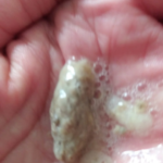
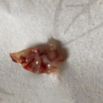
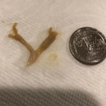
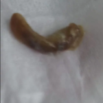
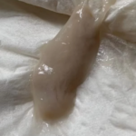
People who have aspergillosis tend to cough up characteristic mucoid plugs often when being given oral steroids. These can take many shapes and forms and be different colours depending on what they contain – some can have hard objects within them, others can be very thick/hard on general consistency.
These can be a helpful sign for a diagnosis, so we thought is would be useful if we collected a small library of images for clinicians to refer to, which we will publish here. Patients could also use them to show to your doctor as examples of the kind of thing they are also coughing up.
Images have helpfully been donated by members of the National Aspergillosis Support UK Facebook group.
Contact us if you have examples you would like us to include.
[/et_pb_text][et_pb_toggle title="Aspergillosis plugs ‘n slugs (Click here to show images)" open_toggle_text_color="#000000" _builder_version="4.18.0" _module_preset="default" title_text_color="#000000" title_font="|600|||||||" global_colors_info="{}" theme_builder_area="post_content"]
[/et_pb_toggle][/et_pb_column][/et_pb_row][/et_pb_section]
Living with CPA and ABPA
[et_pb_section fb_built="1" _builder_version="4.18.0" _module_preset="default" global_colors_info="{}" theme_builder_area="post_content" custom_padding="8px||2px|||"][et_pb_row _builder_version="4.18.0" _module_preset="_initial" custom_margin="-21px|auto||auto||" global_colors_info="{}" theme_builder_area="post_content" custom_padding="40px|0px|40px|0px|true|true"][et_pb_column type="4_4" _builder_version="4.18.0" _module_preset="default" global_colors_info="{}" theme_builder_area="post_content"][et_pb_text _builder_version="4.18.0" _module_preset="default" global_colors_info="{}" theme_builder_area="post_content"]
Gwynedd was formally diagnosed with CPA and ABPA at the National Aspergillosis Centre in 2012. Below she lists some of the symptoms she experiences and what she has found helpful in managing the conditions.
These symptoms fluctuate and can be very insignificant until a flare-up occurs. Then they can be severe enough to alter what I can do in a day.
- Tightening of the chest and or upper airway.
- Inflammation can be felt as heat and a 'zingyness' in my chest.
- Pain and discomfort over my back in my lungs.
Self-help
- A healthy diet, as recommended by the dietetic society or as guided by a consultant or specialist nurse.
- Extra protein where one is underweight.
- Exercise is essential for my mental well-being and helps me with chest clearing.
My local respiratory consultant firmly believes in the benefits of Yoga and slower breathing to help with chest clearance and relaxation, which reduces inflammation and anxiety and aids the immune system.
Anxiety is a side effect of ABPA & CPA as both conditions are debilitating, and fluctuations occur seemingly with no warning. It is not unreasonable to feel anxious about this diagnosis. Treatments help, as do lifestyle changes.
[/et_pb_text][/et_pb_column][/et_pb_row][/et_pb_section]
Ear, Eye and Nail Aspergillus infections
[et_pb_section fb_built="1" _builder_version="4.18.0" _module_preset="default" custom_padding="3px|||||" saved_tabs="all" global_colors_info="{}" theme_builder_area="post_content"][et_pb_row _builder_version="4.18.0" _module_preset="default" global_colors_info="{}" theme_builder_area="post_content"][et_pb_column type="4_4" _builder_version="4.18.0" _module_preset="default" global_colors_info="{}" theme_builder_area="post_content"][et_pb_text _builder_version="4.18.0" _module_preset="default" text_text_color="gcid-7b166f65-c038-497a-80df-cc2daa08dd2b" header_text_color="gcid-606e30fd-4e27-4cd6-9cb8-83dbf2f26467" inline_fonts="ABeeZee,Arial" global_colors_info="{%22gcid-7b166f65-c038-497a-80df-cc2daa08dd2b%22:%91%22text_text_color%22%93,%22gcid-606e30fd-4e27-4cd6-9cb8-83dbf2f26467%22:%91%22header_text_color%22%93}" theme_builder_area="post_content"]
Ear, Eye and Nail Aspergillus infections
[/et_pb_text][et_pb_text _builder_version="4.18.0" _module_preset="default" text_font_size="18px" global_colors_info="{}" theme_builder_area="post_content"]
Otomycosis
Onychomycosis
Fungal Keratitis
[/et_pb_text][/et_pb_column][/et_pb_row][et_pb_row _builder_version="4.18.0" _module_preset="157b2edb-eee9-4384-8648-c0e491cc6118" global_colors_info="{}" theme_builder_area="post_content"][et_pb_column type="4_4" _builder_version="4.18.0" _module_preset="default" global_colors_info="{}" theme_builder_area="post_content"][et_pb_text module_id="otomycosis" _builder_version="4.18.0" _module_preset="default" global_colors_info="{}" theme_builder_area="post_content"]
Otomycosis
Otomycosis is a fungal infection of the ear, and the most frequently encountered fungal infection in ear, nose and throat clinics. The organisms responsible for otomycosis are usually fungi from the environment, most commonly Aspergillus niger. The fungi usually invade tissue that has already been damaged by bacterial infections, physical injury or excess earwax.
[/et_pb_text][et_pb_image src="https://aspergillosis.org/wp-content/uploads/2022/10/Otitis_externa-768x576-1.jpg" title_text="Otitis_externa-768x576" align="center" _builder_version="4.18.0" _module_preset="default" width="50%" global_colors_info="{}" theme_builder_area="post_content"][/et_pb_image][et_pb_text _builder_version="4.18.0" _module_preset="default" global_colors_info="{}" theme_builder_area="post_content"]
Symptoms:
- Itching, irritation, discomfort or pain
- Small amounts of discharge
- A feeling of blockage in the ear
In rare cases, Aspergillus infecting the ear may spread to bone and cartilage, causing a severe and life-threatening disease. This is more frequently caused by Aspergillus fumigatus than Aspergillus niger, and is associated with underlying immunocompromisation, diabetes mellitus or patients on dialysis.
A diagnosis of otomycosis is confirmed by taking debris from the infected ear, culturing it on a special agar plate and using microscopy to establish the causative organism. If the infection is deep, a biopsy should be taken for fungal culture and identification. If there is a suspicion of the infection becoming invasive, CT and MRI scans can be used to see whether the fungi has spread to any other sites.
Treatment involves carefully drying and cleaning the ear canal, using microsuction. Aural syringing should be avoided as it can lead to the infection flaring up in deeper sites of the ear. Depending on how complicated the infection is, you may need to further treat with antifungals applied to the ear. Treatment should continue for 1-3 weeks and oral antifungal therapy is only required if the antifungals applied to the skin do not work, or the condition is invasive.
With good ear canal cleaning and antifungal therapy, otomycosis is usually cured and does not relapse.
Click here for more information on otomycosis
[/et_pb_text][/et_pb_column][/et_pb_row][et_pb_row _builder_version="4.18.0" _module_preset="157b2edb-eee9-4384-8648-c0e491cc6118" custom_margin="60px|auto||auto||" global_colors_info="{}" theme_builder_area="post_content"][et_pb_column type="4_4" _builder_version="4.18.0" _module_preset="default" global_colors_info="{}" theme_builder_area="post_content"][et_pb_text module_id="Onychomycosis" _builder_version="4.18.0" _module_preset="default" global_colors_info="{}" theme_builder_area="post_content"]
Onychomycosis
Onychomycosis is a fungal infection of the nail, most commonly the toenail. Fungal nail infection is common in the general adult population, with a rate of about 5-25% and increasing incidence in elderly people. Onychomycosis makes up about 50% of all nail disease. There are a wide variety of fungi that can onychomycosis, but T. rubrum is responsible for about 80% of cases in the UK. Aspergillus species, amongst many other fungi, can occasionally cause onychomycosis. Some infections are caused by more than one fungus.
[/et_pb_text][et_pb_image src="https://aspergillosis.org/wp-content/uploads/2022/10/Fungal-nail-infection-1.png" title_text="Fungal-nail-infection-1" align="center" _builder_version="4.18.0" _module_preset="default" width="50%" global_colors_info="{}" theme_builder_area="post_content"][/et_pb_image][et_pb_text _builder_version="4.18.0" _module_preset="default" global_colors_info="{}" theme_builder_area="post_content"]
Symptoms of the infection will vary depending on the type of fungus involved, but thickened nails and discoloration are common.
Some of the contributing factors causing this disease are occlusive footwear, extensive water contact with nails, repeated nail trauma, genetic predisposition and concurrent disease, such as diabetes, poor peripheral circulation and HIV infection, as well as other forms of immunosuppression.
Diagnosis of the causative fungus is achieved by scraping the nail (the material under the nail is the most rewarding material). Small pieces of this are then inspected under a microscope and grown on special agar plates to determine the species responsible for the disease.
Treatment depends on the causative species and the severity of the disease. Antifungal cream or ointment applied to the affected nail is effective in some milder cases. Oral antifungal therapy or surgery to remove the nail may be required. Treatment can last from 1 week to 12+ months, depending on the case. Cure is possible, but takes a long time, as the growth of nails is slow.
The nail fold can also become infected – this is called paronychia, and is usually caused by Candida albicans and other Candida species.
Click here for more information on onychomycosis
[/et_pb_text][/et_pb_column][/et_pb_row][et_pb_row _builder_version="4.18.0" _module_preset="157b2edb-eee9-4384-8648-c0e491cc6118" custom_margin="60px|auto||auto||" global_colors_info="{}" theme_builder_area="post_content"][et_pb_column type="4_4" _builder_version="4.18.0" _module_preset="default" global_colors_info="{}" theme_builder_area="post_content"][et_pb_text module_id="fungal-keratitis" _builder_version="4.18.0" _module_preset="default" global_colors_info="{}" theme_builder_area="post_content"]
Fungal Keratitis
Fungal keratitis is a fungal infection of the cornea. The most common causative agents are Aspergillus flavus, Aspergillus fumigatus, Fusarium spp. and Candida albicans, although other fungi can be responsible. Trauma, especially if associated with plant material, is a common antecedent to fungal keratitis. Contact lens fluid contaminated with fungi can also cause fungal keratitis. Other possible risk factors include topical corticosteroids, traditional medicines and higher external temperatures and humidity. Bacterial keratitis is more common in contact lens wearers and the western world, whereas in India and Nepal and some other countries, fungal keratitis is at least as common as bacterial keratitis. There are estimated to be over a million cases of fungal keratitis annually worldwide, mostly in tropical countries.
[/et_pb_text][et_pb_image src="https://aspergillosis.org/wp-content/uploads/2022/10/eyefungal.jpg" title_text="eyefungal" align="center" _builder_version="4.18.0" _module_preset="default" width="50%" global_colors_info="{}" theme_builder_area="post_content"][/et_pb_image][et_pb_text _builder_version="4.18.0" _module_preset="default" global_colors_info="{}" theme_builder_area="post_content"]
ymptoms are usually like other types of keratitis, but perhaps more prolonged in duration (5-10 days):
- eye redness
- pain
- excess tears or other discharge from your eye
- difficulty opening your eyelid because of pain or irritation
- blurred vision
- decreased vision
- sensitivity to light
- a feeling that something is in your eye
The best way to diagnose fungal keratitis is to take a scraping of infective material from the cornea. Any fungal agent in this scraping is then grown on a special agar plate for identification. Along with culturing the organism, microscopy is required due to the wide variety of potential causative fungi.
Antifungals applied directly to the eye in the form of eye drops are essential for the treatment of fungal keratitis. The frequency at which they are administered depends on the severity of the infection. In severe cases this is hourly, and can be reduced in frequency after 1 day as improvement is documented. Topical antifungal therapy has a 60% response rate with retention of vision if keratitis is severe and a 75% response if milder. For severe infections, oral therapy is also advised. The antifungal treatment given depends on the causative species. Therapy is usually continued for at least 14 days. Surgical debridement is essential for severe disease.
Fungal keratitis is associated with a ~5- fold higher risk of subsequent perforation and need for a corneal transplant than bacterial keratitis. Recovery of sight is higher if the diagnosis is made early.
Click here for more information on fungal keratitis
[/et_pb_text][/et_pb_column][/et_pb_row][/et_pb_section]


