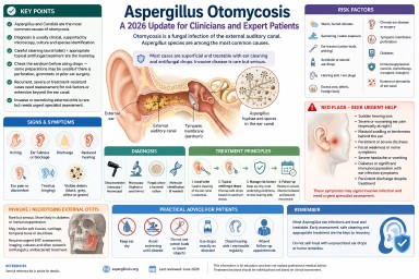
Aspergillus Otomycosis: A 2026 Update for Clinicians and Expert Patients

Article type: Clinical and expert-patient evidence updateIntended audience: Ear, nose and throat clinicians, infectious diseases specialists, medical mycologists, general practitioners, specialist nurses, expert patients and carers.Last reviewed: June 2026
Key points
- Otomycosis is a fungal infection of the external auditory canal. It is often described as fungal otitis externa.
- Aspergillus species and Candida species are the most commonly reported causes.
- Older reports often describe Aspergillus niger as the main cause, but molecular methods show that related black Aspergillus species, including Aspergillus tubingensis, may previously have been misidentified as A. niger.
- Diagnosis is usually clinical, supported where possible by microscopy, fungal culture and species identification.
- Successful treatment usually requires careful cleaning of the ear canal as well as appropriate topical antifungal treatment.
- Before topical treatment is used, clinicians should assess whether the tympanic membrane is intact, because some preparations may be unsafe if there is perforation, grommets, a mastoid cavity or previous ear surgery.
- Most cases are superficial, but recurrent, severe or treatment-resistant disease should prompt reassessment for mixed bacterial infection, diabetes, immunosuppression, hearing aid moulds, foreign body, chronic ear disease or extension beyond the external canal.
- Invasive or necrotising external otitis is rare but serious and requires urgent specialist assessment.
- Sudden hearing loss, facial weakness, severe persistent pain, mastoid swelling or tenderness, neurological symptoms, or infection in a person with diabetes or significant immunosuppression should be treated as red flags.
Contents
- What is otomycosis?
- Aspergillus ear infection is different from lung aspergillosis
- Why Aspergillus matters
- Epidemiology and why cases may be missed
- Causative Aspergillus species
- Risk factors
- Symptoms and clinical features
- Diagnosis
- Differential diagnosis
- Treatment principles
- Recurrent or treatment-resistant otomycosis
- Invasive Aspergillus ear infection and necrotising external otitis
- Practical advice for patients
- Frequently asked questions
- When to seek urgent medical help
- Evidence gaps and uncertainty
- References
What is otomycosis?
Otomycosis is a fungal infection of the external auditory canal, the skin-lined passage leading from the outer ear to the eardrum. It is also called fungal otitis externa. The infection may be acute, subacute or chronic, and it can be mistaken for bacterial otitis externa, eczema, wax, chronic discharge or non-specific inflammation.
In most people, otomycosis is a superficial infection of the outer ear canal. It can be uncomfortable, persistent and prone to recurrence, but it is usually treatable when the ear is examined, cleaned and treated appropriately. Rarely, particularly in people with diabetes, immunosuppression, previous ear surgery or severe persistent symptoms, infection may spread beyond the ear canal and become invasive.
Plain-English summary: most Aspergillus ear infections affect the outer ear canal. They are usually not the same as invasive aspergillosis in the lungs or bloodstream, but they can still need specialist ear care to clear the infection safely.
Aspergillus ear infection is different from lung aspergillosis
Many people searching online for Aspergillus find information about chronic pulmonary aspergillosis, allergic bronchopulmonary aspergillosis or invasive aspergillosis. These are different conditions.
Aspergillus otomycosis usually affects the external ear canal only. It is usually a local fungal infection rather than a whole-body infection. It does not usually mean that a person has invasive aspergillosis, chronic pulmonary aspergillosis or allergic bronchopulmonary aspergillosis.
However, people with existing lung disease, diabetes, immune suppression or complex medical histories should still tell their clinician about these conditions, because they may affect assessment and treatment decisions.
Why Aspergillus matters
Aspergillus is a common environmental mould. Its spores are present in air, dust, soil, compost, decaying vegetation and indoor environments. In the ear canal, fungal growth is favoured when local conditions change. Moisture, trauma to the skin, loss of normal wax protection, altered acidity, previous antibiotic or steroid drops, retained debris and obstruction from hearing aid moulds or ear plugs can all make fungal overgrowth more likely.
In otomycosis, Aspergillus species are among the most frequently reported fungi. They are particularly important because their appearance in the ear can be striking, with black, grey, greenish, yellow-white or fluffy debris. However, appearance alone is not enough for precise species identification.
Epidemiology and why cases may be missed
Reported prevalence varies considerably between studies and geographical regions. Otomycosis is more common in some warm, humid or dusty environments, but it is reported worldwide, including in the United Kingdom. In UK practice, it may be considered particularly in chronic, recurrent or treatment-resistant otitis externa.
Cases may be missed because symptoms overlap with bacterial otitis externa and inflammatory ear conditions. A patient may be treated repeatedly with antibacterial or steroid-containing ear drops before fungal infection is considered. In some cases, antibacterial treatment may suppress bacteria while allowing fungi to overgrow.
A 2025 systematic review highlighted increasing use of molecular methods for species identification, although many published studies still rely primarily on microscopy and culture.
Causative Aspergillus species
Older articles often refer to Aspergillus niger as the dominant Aspergillus species in otomycosis. This remains a useful clinical shorthand, but it is no longer the whole story. Modern molecular identification has shown that black Aspergillus isolates are a complex group and may include species such as Aspergillus tubingensis and other members of the Aspergillus niger complex.
Reported Aspergillus species in otomycosis and fungal otitis externa include:
- Aspergillus niger complex, including related black Aspergillus species
- Aspergillus tubingensis
- Aspergillus flavus
- Aspergillus fumigatus
- Aspergillus terreus complex, reported occasionally in superficial infection series but apparently less common than the Aspergillus niger complex or Aspergillus flavus
Clinical relevance: species-level identification is not always needed for straightforward cases that respond to topical treatment. It becomes more important in recurrent, invasive, immunocompromised or treatment-resistant infection, or where systemic antifungal treatment is being considered.
Risk factors
Otomycosis usually develops when the normal protective environment of the ear canal is disrupted. Important risk factors include:
- warm, humid or dusty environments
- frequent swimming or repeated water exposure
- use of cotton buds, ear picking or other trauma to the ear canal
- previous or repeated antibacterial ear drops
- topical steroid use in the ear
- hearing aids, ear plugs or occlusive moulds
- excess wax, retained debris or foreign material
- chronic otitis externa or chronic otitis media
- previous ear surgery or mastoid cavity
- tympanic membrane perforation
- diabetes, especially if poorly controlled
- immunosuppression, including chemotherapy, transplant medicines, prolonged high-dose corticosteroids and some biological therapies
- skin conditions affecting the ear canal, such as eczema or seborrhoeic dermatitis
Plain-English summary: fungi grow more easily when the ear canal is damp, damaged, blocked, repeatedly treated with antibiotics, or when a person’s immune defences are reduced.
Symptoms and clinical features
Symptoms vary. Some patients have mild itching and fullness, while others have marked discomfort, discharge or hearing loss. Common symptoms include:
- itching in the ear
- ear fullness or blockage
- discharge from the ear
- reduced hearing, often due to debris blocking the canal
- ear discomfort or pain
- tinnitus or ringing in the ear
- scaling, inflammation or visible debris in the ear canal
Severe pain, persistent night pain, swelling around the ear, tenderness or swelling over the mastoid bone behind the ear, fever, persistent or severe dizziness, facial weakness, severe headache, cranial nerve symptoms or symptoms in a person with diabetes or immunosuppression should raise concern for more serious disease.
Sudden hearing loss should be treated as a medical emergency and assessed urgently, regardless of whether otomycosis is suspected.
Diagnosis
Clinical examination
Diagnosis is often suspected by otoscopic or microscopic examination of the ear canal. Typical findings may include fungal debris, spores, hyphae, wet or dry masses, inflammation, scaling, discharge and obstruction. Black, grey, white, yellow-green or fluffy material may be seen, but visual appearance does not reliably identify the species.
Assessment should include:
- extent of external canal inflammation
- presence of fungal debris or discharge
- degree of canal obstruction
- condition of the tympanic membrane
- evidence of perforation, grommets, mastoid cavity or prior surgery
- features suggesting bacterial co-infection
- signs of spread beyond the external canal
Microscopy and fungal culture
Microscopy can demonstrate fungal hyphae or yeast forms. Culture can identify whether Aspergillus, Candida or another fungus is present. Bacterial culture may also be useful if mixed infection is suspected.
Culture is especially helpful when disease is recurrent, severe, atypical, invasive, not responding to standard treatment, or occurring in a person with diabetes, immunosuppression or previous ear surgery.
Molecular identification
Molecular methods can identify Aspergillus species more accurately than morphology alone. This matters because older culture-based reports may have grouped several black Aspergillus species under Aspergillus niger. Molecular identification may also help in epidemiological studies and difficult clinical cases.
For routine mild otomycosis, molecular identification is not always necessary. For persistent, recurrent or invasive disease, species-level identification and antifungal susceptibility testing may help guide treatment.
Antifungal susceptibility testing
Antifungal susceptibility testing is not required for every simple case. It should be considered when there is treatment failure, recurrent disease, unusual species, invasive infection or planned systemic antifungal therapy. Susceptibility patterns may vary between Aspergillus species, although resistance testing is not routinely required for uncomplicated superficial disease.
When to consider imaging
Imaging is not needed for uncomplicated superficial otomycosis. It may be required if there is concern about necrotising external otitis, skull base osteomyelitis, mastoid involvement, middle ear extension, cranial nerve involvement or deep tissue spread. Imaging decisions should be made by ear, nose and throat, infectious diseases, radiology and microbiology or mycology specialists as appropriate.
Differential diagnosis
Conditions that can resemble Aspergillus otomycosis include:
- bacterial otitis externa
- chronic otitis externa
- eczema or dermatitis of the ear canal
- impacted wax
- foreign body
- chronic suppurative otitis media
- cholesteatoma
- otitis media with perforation
- necrotising external otitis
- malignancy of the external canal or temporal bone, rarely
Failure to respond to usual antibacterial treatment should prompt reconsideration of the diagnosis rather than repeated courses of the same treatment.
Treatment principles
Treatment depends on severity, tympanic membrane status, immune status, recurrence, species where known, and whether infection is limited to the outer ear canal. Local ear, nose and throat practice and local antimicrobial guidance should be followed.
1. Aural toilet and debridement
Careful cleaning of the ear canal is central to management. Fungal debris can block the canal, protect organisms from topical treatment and contribute to hearing loss. Aural toilet, often using microsuction or careful debridement, is usually best performed by a trained clinician using appropriate equipment, especially if the canal is swollen, painful, obstructed, or if the tympanic membrane cannot be seen.
Patients should not attempt deep cleaning with cotton buds, hair grips, ear candles or improvised tools. These can damage the ear canal, push debris deeper, worsen inflammation or perforate the eardrum.
Ear irrigation is not usually the preferred approach for suspected otomycosis, because retained moisture may encourage fungal growth and because irrigation may be unsafe if there is tympanic membrane perforation, grommets or previous ear surgery.
2. Topical antifungal treatment
Most uncomplicated otomycosis is treated with topical antifungal therapy after cleaning. Reported agents include clotrimazole, miconazole, nystatin, ciclopirox and other locally used preparations. Different countries and ear, nose and throat services use different formulations, and the evidence does not clearly establish one universally superior topical agent for all cases.
Choice of topical treatment should consider:
- likely organism: mould versus yeast
- whether the tympanic membrane is intact
- previous ear surgery, mastoid cavity or grommets
- local formulary and ear, nose and throat practice
- potential ototoxicity
- patient tolerance and adherence
- whether the canal needs repeat cleaning
3. Tympanic membrane safety
Assessment of the eardrum is important. Some topical agents, antiseptics, acidic preparations or combination drops may be unsafe if the tympanic membrane is perforated or if there are grommets, a mastoid cavity or previous ear surgery. If the eardrum cannot be visualised, treatment should be chosen cautiously and specialist ear, nose and throat advice may be needed.
Important safety point
Patients should not put unprescribed antifungal, antiseptic, acidic, oil-based or herbal preparations into the ear. This is especially important if there is discharge, severe pain, previous ear surgery, grommets, suspected perforated eardrum, sudden hearing loss, dizziness or facial weakness.
4. Mixed bacterial and fungal infection
Mixed infection can occur. If bacterial infection is suspected, bacterial culture and targeted antibacterial treatment may be needed. However, repeated broad-spectrum antibacterial drops without reassessment may encourage fungal overgrowth and delay recognition of otomycosis.
5. Hearing aids, earmoulds and ear plugs
Hearing aids, earmoulds and ear plugs can trap moisture and debris in the ear canal. They may also act as a continuing source of irritation or contamination if not cleaned properly. In recurrent otomycosis, hearing aids and moulds should be reviewed, cleaned according to manufacturer guidance and, where appropriate, assessed by audiology or ear, nose and throat services.
6. Diabetes and immune suppression
People with diabetes or significant immune suppression need particular care, because severe or invasive external ear infection is more likely in these groups. Optimising glycaemic control may help reduce the risk of severe or recurrent infection. Clinicians should consider whether persistent or severe otitis externa in a person with diabetes could represent necrotising external otitis.
7. Systemic antifungal treatment
Systemic antifungal treatment is not usually required for uncomplicated superficial otomycosis. It may be considered in selected situations, including:
- invasive or necrotising external otitis
- extension into the middle ear, mastoid, skull base or surrounding tissues
- severe infection in an immunocompromised patient
- persistent disease where topical therapy is not possible, unsafe or ineffective
- cases requiring multidisciplinary ear, nose and throat, infectious diseases and mycology input
Systemic antifungal choice should be guided by species identification, susceptibility testing, site and extent of infection, drug interactions, renal and liver function, and local specialist advice. Azole antifungals such as itraconazole, voriconazole, posaconazole and isavuconazole have important drug interactions and monitoring requirements.
Recurrent or treatment-resistant otomycosis
Recurrent otomycosis is common in some series. Recurrence does not always mean that the original treatment was wrong; it may reflect ongoing risk factors or incomplete clearance of debris.
When otomycosis recurs or fails to respond, clinicians should reassess:
- Was the ear canal adequately cleaned?
- Can the tympanic membrane be seen?
- Is there perforation, chronic otitis media or mastoid cavity disease?
- Is there a hearing aid mould, ear plug or foreign body contributing?
- Is the patient using cotton buds or self-cleaning the ear?
- Is there persistent water exposure?
- Is there eczema, dermatitis or another inflammatory ear condition?
- Is there mixed bacterial and fungal infection?
- Has microscopy or culture confirmed the organism?
- Is species-level identification or susceptibility testing needed?
- Is the patient diabetic or immunocompromised?
- Is there severe pain, mastoid tenderness or evidence of deeper spread?
Invasive Aspergillus ear infection and necrotising external otitis
Most Aspergillus otomycosis is superficial. However, fungal infection of the external ear can rarely become invasive, particularly in people with diabetes, immunosuppression, severe chronic ear disease or previous ear surgery.
Necrotising external otitis, historically called malignant external otitis, is a severe infection that may involve soft tissues, cartilage, temporal bone or skull base. It is most often bacterial, especially due to Pseudomonas aeruginosa, but fungal cases including Aspergillus and Candida are reported. Fungal necrotising external otitis can be difficult to diagnose and may require prolonged treatment.
Features that should raise concern include:
- severe, persistent or worsening ear pain, especially at night
- pain out of proportion to examination findings
- persistent discharge despite appropriate treatment
- granulation tissue in the ear canal
- facial weakness or other cranial nerve symptoms
- persistent, severe or worsening dizziness, particularly with severe pain, hearing loss or neurological symptoms
- severe headache or neurological symptoms
- swelling, redness or cellulitis around the ear
- tenderness, swelling or redness over the mastoid bone behind the ear
- diabetes, especially if poorly controlled
- immunosuppression
- evidence of mastoid, middle ear or skull base involvement
Suspected invasive or necrotising disease needs urgent ear, nose and throat assessment, microbiological sampling, imaging and specialist antimicrobial or antifungal planning. This is not a condition for self-treatment.
Practical advice for patients and expert patients
Patients can support recovery by protecting the ear canal while treatment is underway. Advice should be individualised by the treating clinician, but common measures include:
- keep the affected ear dry unless advised otherwise
- avoid swimming until cleared by a clinician
- do not use cotton buds or insert objects into the ear
- use prescribed drops exactly as directed
- attend follow-up if symptoms persist, because repeat cleaning may be needed
- tell the clinician about diabetes, immune problems, steroid use, biological therapies, chemotherapy, transplant medicines or previous ear surgery
- ask whether hearing aids, earmoulds or ear plugs need cleaning or review
- report severe pain, dizziness, facial weakness, worsening hearing loss or persistent discharge urgently
Important: online images of invasive aspergillosis can be frightening and often do not represent ordinary otomycosis. Most Aspergillus ear infections are local infections of the outer ear canal. The key is proper assessment, safe cleaning and appropriate treatment.
Frequently asked questions
Is Aspergillus otomycosis rare?
It depends on the setting. Otomycosis is common in some warm and humid regions but is less commonly recognised in many UK clinics. Aspergillus otomycosis may be unfamiliar to clinicians who do not often see fungal ear disease.
Is it the same as invasive aspergillosis?
Usually, no. Most Aspergillus otomycosis is a superficial infection of the outer ear canal. Invasive disease is rare but important, especially in people with diabetes, immunosuppression, severe pain or persistent infection.
Can an ear infection cause symptoms elsewhere in the body?
Otomycosis itself would not usually cause breathlessness, chest symptoms or systemic illness. If a person with suspected otomycosis is also breathless, has chest pain, fever, low oxygen levels or feels very unwell, those symptoms should be assessed separately and promptly.
Can it affect hearing?
Yes. Hearing may be reduced if the ear canal is blocked by fungal debris, discharge or swelling. Hearing often improves when the canal is cleaned and inflammation settles. Sudden hearing loss should be treated as a medical emergency and assessed urgently.
Should every case be cultured?
Not necessarily. Straightforward cases may be treated clinically. Culture and microscopy are more useful when symptoms are recurrent, severe, atypical, treatment-resistant, associated with perforation or surgery, or occur in an immunocompromised patient.
Are antifungal ear drops enough?
Often, topical treatment is effective, but drops may not work well if the ear canal is blocked with debris. Cleaning the canal is often as important as the antifungal medicine itself.
Can patients buy something and treat it themselves?
This is not recommended. Ear symptoms can have several causes, and some substances may be unsafe if the eardrum is perforated. Patients should seek clinical assessment before putting unprescribed antifungal, antiseptic, acidic, herbal or oil-based preparations into the ear.
Why does otomycosis come back?
Recurrence can happen if the canal remains damp, debris persists, the ear is repeatedly traumatised, a hearing aid mould traps moisture, eczema is present, antibiotics are repeatedly used, or an underlying condition such as diabetes or immune suppression is not recognised.
When to seek urgent medical help
Patients should seek urgent medical advice if they have:
- severe or worsening ear pain
- pain that wakes them at night
- swelling, redness or tenderness around the ear
- tenderness, swelling or redness over the mastoid bone behind the ear
- persistent discharge despite treatment
- sudden hearing loss or rapidly worsening hearing
- persistent, severe or worsening dizziness
- severe headache or vomiting
- facial weakness or changes in facial movement
- confusion, fever or feeling very unwell
- diabetes, immune suppression or recent chemotherapy or transplant treatment with ear infection symptoms
- previous ear surgery, grommets or known eardrum perforation with new discharge or pain
These symptoms do not prove invasive infection, but they should be assessed promptly.
Evidence gaps and uncertainty
The evidence base for otomycosis is improving but remains limited. Many studies are single-centre observational series from regions where otomycosis is more common. Treatment studies vary in diagnostic criteria, topical agents, follow-up duration and whether cleaning was standardised. There is no single universally accepted treatment regimen for all cases.
Important gaps include:
- limited high-quality randomised trials comparing topical antifungal treatments
- variable reporting of tympanic membrane status and previous ear surgery
- inconsistent use of fungal culture, molecular identification and susceptibility testing
- limited data on recurrence prevention
- limited evidence specific to UK practice
- uncertainty about the best systemic antifungal approach in rare invasive cases
For these reasons, this article should support but not replace specialist clinical judgement.
References
- Nazari T, Peymaeei F, Ghazi Mirsaid R, et al. Otomycosis: a systematic review and meta-analysis of prevalence and causative agents in the era of molecular diagnostics. BMC Infectious Diseases. 2025;25(1). doi: 10.1186/s12879-025-10954-y. PubMed
- Bojanović M, Stalević M, Arsić-Arsenijević V, et al. Etiology, Predisposing Factors, Clinical Features and Diagnostic Procedure of Otomycosis: A Literature Review. Journal of Fungi. 2023;9(6):662. doi: 10.3390/jof9060662. PubMed
- Wiegand S, Berner R, Schneider A, Lundershausen E, Dietz A. Otitis Externa: Investigation and Evidence-Based Treatment. Deutsches Ärzteblatt International. 2019. doi: 10.3238/arztebl.2019.0224. PubMed
- Koltsidopoulos P, Skoulakis C. Otomycosis With Tympanic Membrane Perforation: A Review of the Literature. Ear, Nose & Throat Journal. 2019;99(8):518–521. doi: 10.1177/0145561319851499. PubMed
- Mtibaa L, Halwani C, El Hamdi M, et al. A retrospective study of 43 cases of fungal malignant external otitis. Pan African Medical Journal. 2022;41. doi: 10.11604/pamj.2022.41.287.29585. PubMed
- Szigeti G, Sedaghati E, Mahmoudabadi AZ, et al. Species assignment and antifungal susceptibilities of black aspergilli recovered from otomycosis cases in Iran. Mycoses. 2011;55(4):333–338. doi: 10.1111/j.1439-0507.2011.02103.x. PubMed
- Jimenez-Garcia L, Celis-Aguilar E, Díaz-Pavón G, et al. Efficacy of topical clotrimazole vs. topical tolnaftate in the treatment of otomycosis: a randomized controlled clinical trial. Brazilian Journal of Otorhinolaryngology. 2020;86(3):300–307. doi: 10.1016/j.bjorl.2018.12.007. PubMed
- Mofatteh MR, Naseripour Yazdi Z, Yousefi M, et al. Comparison of the recovery rate of otomycosis using betadine and clotrimazole topical treatment. Brazilian Journal of Otorhinolaryngology. 2018;84(4):404–409. doi: 10.1016/j.bjorl.2017.04.004. PubMed
- Mao C, Li A, Hu J, et al. Efficient and accurate diagnosis of otomycosis using an ensemble deep-learning model. Frontiers in Molecular Biosciences. 2022;9:951432. doi: 10.3389/fmolb.2022.951432. PubMed
- Feng Y, Zhang Z, Fang W, et al. Profiling Drug Susceptibility and Species Identification of Aspergillus Isolates From Patients With Superficial Infection. Mycoses. 2025;68(4). doi: 10.1111/myc.70059. PubMed
- NICE Clinical Knowledge Summaries. Otitis externa. NICE CKS
Author and review information
Article type: Clinical and expert-patient evidence update
Intended audience: Ear, nose and throat clinicians, infectious diseases specialists, medical mycologists, general practitioners, specialist nurses, patients and carers seeking detailed information.
Last reviewed: June 2026
Review note: This article is for information and education. It should not replace assessment by a qualified clinician. Treatment choices depend on examination findings, tympanic membrane status, culture results where available, medical history, immune status and local prescribing guidance.
```
Sinusitis in Patients with ABPA
When to suspect it, when to investigate, and when to refer
Why this matters
Patients with allergic bronchopulmonary aspergillosis (ABPA) are usually managed as having a lung disease. Diagnosis, monitoring, and treatment focus appropriately on the chest, immunology, and asthma control.
However, ABPA occurs within a single continuous airway, extending from the nose and sinuses to the lungs. Disease in the upper airway can coexist with, exacerbate, or complicate lower airway inflammation — yet sinus disease is not routinely assessed in ABPA care pathways.
This article outlines:
-
What is known about sinus disease in this context
-
Which symptoms should raise suspicion
-
When investigation or ENT referral should be considered
-
What GPs and non-specialists can reasonably do
The united airway: a brief reminder
The upper and lower airways share:
-
Type 2 (eosinophilic) inflammation
-
Immunoglobulin E–mediated immune responses
-
Common triggers, including allergens and fungi
Chronic rhinosinusitis is common in asthma and severe asthma, and treatment of sinus disease can improve lower airway outcomes in some patients.
ABPA sits within this same inflammatory spectrum, even though its management is lung-centred.
Sinus disease in ABPA: what is (and isn’t) known
What we know
-
Chronic rhinosinusitis is common in patients with asthma and severe asthma
-
Sinus disease may be symptomatic or relatively silent
-
ABPA guidelines do not mandate routine ENT review or sinus imaging
-
ENT involvement, therefore, varies widely between centres
What we do not know
-
Whether routine ENT assessment improves ABPA outcomes
-
Which ABPA patients benefit most from sinus intervention
-
The optimal timing for ENT referral in ABPA
As a result, clinical judgement remains central.
Symptoms that should prompt consideration of sinus disease
Sinusitis in ABPA patients does not always present with classic “blocked nose and facial pain”.
Key symptoms include:
Common but often overlooked
-
Persistent post-nasal drip
-
Foul, bitter, metallic, or “infected” taste in the mouth
-
Throat clearing, chronic cough
-
Thick or sticky mucus sensation
-
Symptoms are worse on waking or lying flat
More typical sinonasal features
-
Nasal blockage or congestion
-
Facial pressure or fullness
-
Reduced or altered sense of smell
-
Nasal crusting or discharge
Contextual clues
-
Poor durability of response to steroids or antifungals
-
Recurrent “flares” without clear chest triggers
-
Coexisting severe asthma or nasal polyps
-
Symptoms are worse in damp or mould-affected housing
A persistent foul taste in the mouth is a recognised symptom of chronic sinus disease, usually due to post-nasal drainage of inflamed secretions.
Damp homes and sinus disease
Living in damp or mould-affected environments is associated with:
-
Higher rates of chronic rhinosinusitis
-
Upper airway irritation and inflammation
-
Allergic sensitisation to fungal spores
In most cases, this results in inflammatory or allergic sinusitis, not invasive fungal infection.
Fungal involvement may act as an immune trigger, even when not labelled as “fungal sinusitis”.
Fungal sinusitis: rare vs under-recognised
It is important to distinguish between entities:
| Type | Frequency | Key point |
|---|---|---|
| Invasive fungal sinusitis | Rare | Usually immunocompromised; dramatic presentation |
| Fungal ball (mycetoma) | Uncommon | Usually obvious on CT |
| Allergic fungal rhinosinusitis | Likely under-recognised | Requires active suspicion |
Allergic fungal rhinosinusitis overlaps biologically with ABPA:
-
IgE-mediated
-
Eosinophilic inflammation
-
Thick allergic mucin
It is not routinely sought, so it may be under-diagnosed in at-risk groups.
What GPs and non-specialists can reasonably do
1. Take upper airway symptoms seriously
Especially in ABPA or severe asthma patients with:
-
Persistent post-nasal symptoms
-
Foul taste
-
Recurrent unexplained deterioration
2. Examine the nose and throat
-
Look for polyps, discharge, and crusting
-
Note mouth breathing or altered voice quality
-
Check dentition (to exclude dental causes)
3. Consider imaging when symptoms persist
-
CT sinuses (not plain X-ray) is the imaging of choice
-
Particularly appropriate if symptoms last >8–12 weeks or recur
4. Refer to ENT when:
-
Symptoms are persistent or progressive
-
CT shows significant sinus disease
-
There is a poor response to standard medical therapy
-
There is diagnostic uncertainty
Referral does not imply surgery — ENT input may be diagnostic or medical.
What this article is not saying
-
It does not suggest that all ABPA patients need an ENT referral
-
It does not claim that sinus treatment improves ABPA outcomes
-
It does not override existing guidelines
It does suggest that earlier consideration of the upper airway is reasonable in selected patients.
Key take-home points for clinicians
-
The airway functions as a single inflammatory system
-
Sinus disease may be subtle, under-reported, or atypical
-
A foul taste in the mouth is a meaningful symptom
-
Damp or mould exposure increases sinus disease risk
-
ENT referral is appropriate when symptoms persist or recur
-
Evidence gaps remain — but clinical vigilance is justified
In summary
ABPA is managed as a lung disease, but patients live with a whole airway.
Recognising when sinus disease may be contributing can help explain persistent symptoms and guide appropriate referral — without over-investigation or over-treatment.
⭐ Aspergillus Bronchitis: The Overlooked Condition Hiding in Plain Sight
Estimated prevalence 1–2% in bronchiectasis and chronic airway disease clinics.
Aspergillus Bronchitis (AB) is a chronic, symptomatic infection of the airways caused by Aspergillus species in people with underlying lung disease. It sits between simple colonisation and chronic pulmonary aspergillosis (CPA), and is frequently overlooked or mislabelled as “recurrent infection,” “post-viral symptoms,” or uncontrolled bronchiectasis.
Unlike CPA, Aspergillus Bronchitis does not require cavities or major structural destruction — which makes it both easier to miss and surprisingly common among people with chronic airway disease.
When recognised and treated (usually with antifungal therapy for several months), symptoms often improve significantly. But because awareness remains low, most patients cycle through unnecessary antibiotics, repeated exacerbations, and worsening airway disease before the real cause is identified.
⭐ What Exactly Is Aspergillus Bronchitis?
Aspergillus Bronchitis is defined by:
-
chronic productive cough
-
sputum growing Aspergillus species repeatedly
-
airway inflammation
-
symptoms lasting over 3 months
-
underlying airway disease (bronchiectasis, CF, COPD, prior TB, ABPA)
-
response to antifungal therapy
Unlike ABPA:
-
there is no allergic response,
-
IgE is usually normal,
-
eosinophils are normal or mildly elevated.
Unlike CPA:
-
there are no cavities on imaging,
-
IgG may be normal or only slightly elevated,
-
disease is confined to the airways, not lung tissue.
This places AB in a “grey zone” — often invisible unless specifically looked for.
⭐ Why Aspergillus Bronchitis Is Missed
1. Symptoms mimic common chronic airway disease
Typical AB symptoms include:
-
daily productive cough
-
worsening sputum thickness
-
breathlessness
-
fatigue
-
repeated “chest infections”
-
slow-to-clear mucus
-
crackles or wheeze
These resemble:
-
bronchiectasis exacerbations
-
COPD flare-ups
-
chronic infection with Pseudomonas or NTM
-
post-viral cough
-
uncontrolled asthma
Without fungal awareness, clinicians default to bacterial explanations.
2. Sputum grows multiple organisms — Aspergillus is dismissed
In bronchiectasis, sputum frequently grows:
-
Haemophilus
-
Pseudomonas
-
Staphylococcus
-
Streptococcus
-
NTM
When Aspergillus appears, it’s often labelled:
-
“colonisation”
-
“contaminant”
-
“not clinically relevant”
But repeated isolation with persistent symptoms is highly suggestive of AB.
3. IgE/IgG results may be normal
Many clinicians expect high IgE or IgG to “confirm Aspergillus disease.”
But in Aspergillus Bronchitis:
-
IgE is usually normal
-
IgG can be normal or borderline
This leads to false reassurance.
4. Radiology rarely shows overt features
CT scans in AB may show:
-
mucus plugging
-
mild bronchial wall thickening
-
small nodules
-
progression of bronchiectasis
But they do not show the cavities of CPA or classic features of ABPA.
Therefore radiologists often report scans as “no significant change” or “stable bronchiectasis.”
5. Antibiotics appear to help — temporarily
Patients often improve slightly with:
-
amoxicillin
-
doxycycline
-
macrolides
-
ciprofloxacin
This gives clinicians the impression of bacterial disease, but symptoms soon return.
6. Lack of awareness
Many specialists (even in respiratory clinics) are unaware that Aspergillus Bronchitis:
-
exists as a distinct clinical entity
-
can be disabling
-
responds to antifungals
-
predicts progression to CPA if untreated
This leads to significant diagnostic delay.
⭐ Who Is at Highest Risk?
1. Bronchiectasis
The largest risk group.
Aspergillus Bronchitis may account for 1–2% of all bronchiectasis patients, and up to 5–10% in severe or frequent exacerbator groups.
2. Cystic Fibrosis (CF)
These patients frequently grow Aspergillus but not all have ABPA — some have Aspergillus Bronchitis.
3. COPD and chronic productive cough
Especially those with:
-
frequent mucus plugging
-
repeated “infective exacerbations”
-
progressive sputum production
4. Post-TB airway damage
Chronic airway deformity, scarring, and bronchiectasis from old TB predispose to fungal infection.
5. Post-COVID structural disease
A new and growing risk group, especially after prolonged ICU ventilation.
6. ABPA patients
Some patients develop Aspergillus Bronchitis during steroid-dominated treatment or after stopping antifungals.
⭐ Which Specialities Need Greater Awareness?
-
Respiratory medicine
(especially bronchiectasis clinicians and severe asthma teams) -
Infectious Diseases
(frequent respiratory presentations with chronic airway infection) -
Radiology
(to recognise subtle but progressive airway changes) -
Primary care
(“recurrent chest infection” or “persistent cough” patients) -
Physiotherapy & airway clearance teams
(excessive sputum with fungal elements) -
Cystic Fibrosis services
The National Aspergillosis Centre is the ideal referral destination when diagnosis is uncertain or symptoms persist despite typical management.
⭐ Red Flags Suggesting Aspergillus Bronchitis
1. Chronic (>3 months) productive cough + repeated Aspergillus in sputum
Even 2 positive sputums in the right clinical context should raise suspicion.
2. Bronchiectasis patient not improving on repeated antibiotics
3. Thick, tenacious mucus with black, grey, or brown plugs
4. Worsening CT bronchiectasis or mucus plugging
5. Absence of features typical for ABPA (normal IgE, no fleeting infiltrates)
6. Asthma or COPD patient with new persistent sputum
7. Partial response to antibiotics but rapid relapse
8. Unexplained fatigue and breathlessness in someone with airway disease
⭐ The Cost of Missed Aspergillus Bronchitis
If AB is not recognised early, consequences include:
-
repeated exacerbations
-
accelerating bronchiectasis
-
long-term airway damage
-
chronic inflammation
-
steroid overuse
-
unnecessary antibiotics
-
repeated hospitalisations
-
progression to CPA in some patients
For health systems, missed diagnosis leads to:
-
higher admission rates
-
inappropriate long-term antibiotic use
-
avoidable CT scans and investigations
-
greater long-term burden of CPA
But antifungal therapy — when appropriately used — can offer marked symptom improvement and reduce exacerbation frequency.
⭐ Conclusion
Aspergillus Bronchitis is a distinct, treatable form of chronic airway disease seen in people with bronchiectasis, asthma, COPD, CF, and post-TB lung damage. Yet lack of awareness means many patients are repeatedly misdiagnosed with bacterial infections or unexplained chronic cough.
Recognising red flags, reviewing sputum results carefully, and considering antifungal therapy can dramatically improve outcomes. Early referral to specialist centres such as the National Aspergillosis Centre is recommended for complex cases or uncertain diagnosis.
Early identification prevents airway deterioration — and reduces the likelihood of progression to CPA.
🌿 Aspergillosis in the Sinuses (Allergic Fungal Rhinosinusitis – AFRS)
It’s quite possible for Aspergillus to affect both the lungs and the sinuses.
The sinuses are small air-filled spaces behind the nose, eyes, and cheeks that normally drain mucus freely. When Aspergillus spores become trapped there, they can trigger an allergic or inflammatory reaction — rather like ABPA in the lungs.
This allergic form is called Allergic Fungal Rhinosinusitis (AFRS).
It isn’t a contagious infection — it’s an overreaction of the immune system to fungal spores. Over time, it can lead to thick mucus, nasal blockage, and sometimes nasal polyps.
🩵 Common symptoms
-
Persistent nasal congestion or blockage
-
Thick or sticky mucus (sometimes with brown or dark flecks)
-
Reduced or lost sense of smell
-
Facial pressure, pain, or fullness (especially around the eyes or cheeks)
-
Post-nasal drip (mucus running down the throat)
-
Fatigue or worsening asthma symptoms
🔬 Diagnosis
-
CT scan of the sinuses – shows thickened or blocked areas
-
Nasal endoscopy – a tiny camera used to look inside
-
Fungal culture or microscopy from mucus samples
-
Blood tests – sometimes show raised total IgE or Aspergillus-specific IgE
💊 Treatment
-
Topical nasal steroids (sprays or rinses) or short courses of oral steroids to reduce inflammation
-
Saline rinses to help keep the sinuses clear
-
ENT surgery if sinuses are blocked or filled with thick fungal debris
-
Antifungal medication in some cases, especially if fungal growth is confirmed
🌸 The Main Types of Aspergillus Sinus Disease
There are several recognised types of sinus aspergillosis. Most people with ABPA or asthma experience only the allergic form (AFRS).
1️⃣ Allergic Fungal Rhinosinusitis (AFRS)
-
Caused by an allergic reaction to Aspergillus
-
Leads to inflammation, thick mucus, and polyps
-
Non-invasive – the fungus stays on the surface
-
Managed with steroids, nasal rinses, and sometimes surgery
✅ This is the type most relevant for ABPA patients.
2️⃣ Fungal Ball (Mycetoma)
-
A clump of fungus (usually A. fumigatus) in a single sinus, often the cheek (maxillary) sinus
-
Common in otherwise healthy people
-
Causes chronic congestion or facial pain
-
Treated surgically – antifungals rarely needed
-
Not allergic and not invasive
3️⃣ Invasive Aspergillus Sinusitis
-
Rare, seen mostly in people with severely weakened immunity (e.g., chemotherapy, bone marrow transplant, uncontrolled diabetes)
-
The fungus invades surrounding tissue and blood vessels
-
Causes severe facial pain, swelling, fever, sometimes affecting the eyes
-
Needs urgent treatment with antifungal drugs and surgery
⚠️ Very rare in people with ABPA or CPA.
🤝 Specialist care
If you have lung aspergillosis (such as ABPA or CPA) and start noticing more nasal congestion, sinus pressure, or post-nasal drip, it’s sensible to mention it to your respiratory or mycology team.
At the National Aspergillosis Centre (NAC), sinus disease is often co-managed by ENT surgeons, respiratory physicians, and mycology specialists, ensuring coordinated care.
With the right combination of treatments — and early recognition of symptoms — most people find their sinus symptoms improve, and controlling sinus inflammation can even help with overall breathing and energy.
Other forms of aspergillosis: Aspergillus Pneumonia (Community-Acquired Aspergillus Lung Infection)
What is it?
Aspergillus pneumonia is a rare but serious lung infection caused by breathing in spores of the Aspergillus mould (most often Aspergillus fumigatus). Unlike allergic conditions such as Allergic Bronchopulmonary Aspergillosis (ABPA) or Aspergillus bronchitis, which affect the airways, Aspergillus pneumonia occurs when the fungus actually invades lung tissue. This makes it a more dangerous condition.
How do people catch it?
Most cases are acquired in the community (outside hospital).
You may be at higher risk if you have:
-
A weakened immune system (chemotherapy, transplant, high-dose steroids, uncontrolled diabetes).
-
Chronic lung disease such as COPD or emphysema.
-
A very heavy exposure to fungal spores (compost heaps, rotting bark, mulch, or farming dust).
Almost all cases are due to Aspergillus fumigatus, though other species like A. flavus have also been reported.
How common is it?
-
Aspergillus pneumonia is uncommon, despite Aspergillus spores being everywhere in the environment.
-
It is most often seen in people with weak immune defences, long-term lung disease, or very high exposure.
-
Because it often looks like ordinary bacterial or viral pneumonia, it can be missed or diagnosed late.
Symptoms
The illness may start like a regular chest infection:
-
Cough (dry or with sputum)
-
Shortness of breath
-
Fever or chills
-
Chest pain
-
Fatigue
-
Sometimes coughing up blood
It may progress:
-
Slowly over weeks, with cavities (holes) forming in the lungs.
-
Very quickly, especially after flu, COVID-19, or high spore exposure.
How does it differ from other Aspergillus conditions?
| Condition | What’s happening | Who gets it | Key signs |
|---|---|---|---|
| Aspergillus Pneumonia | Fungus invades lung tissue (serious infection) | Immunocompromised patients, COPD, heavy spore exposure | Pneumonia-like illness: fever, cough, breathlessness, chest pain |
| ABPA | Allergy to Aspergillus spores causes airway inflammation | People with asthma or cystic fibrosis | Wheeze, thick mucus plugs, recurrent asthma attacks |
| Aspergillus Bronchitis | Fungus grows in widened/damaged airways without invading tissue | People with bronchiectasis or chronic airway disease | Chronic cough, mucus, sometimes blood streaks |
👉 In short:
-
Pneumonia = infection inside lung tissue (dangerous, urgent).
-
ABPA = allergic reaction in the lungs.
-
Bronchitis = long-term airway infection.
Is it a type of invasive aspergillosis?
Yes. Aspergillus pneumonia is considered a form of invasive aspergillosis because the fungus invades lung tissue:
-
Acute/severe form: fast, aggressive illness in very vulnerable people (immunocompromised, post-viral, heavy spore exposure).
-
Subacute or “necrotising” form: slower, smouldering infection in people with COPD, diabetes, or long-term steroids, often with cavities.
👉 It is not mild like ABPA or bronchitis — it requires antifungal treatment.
Diagnosis
Doctors may use:
-
Chest X-ray or CT scan – patches, cavities, or diffuse shadowing.
-
Sputum or bronchoscopy samples – to detect Aspergillus in culture or under the microscope.
-
Blood tests – for Aspergillus antibodies, or sometimes antigen (galactomannan).
Treatment
-
Antifungal medicines are the main treatment (voriconazole is most common; sometimes itraconazole or posaconazole).
-
Corticosteroids may be added in severe “pneumonitis-type” illness with overwhelming inflammation.
If treatment begins early, many people respond well. If diagnosis is delayed, the illness can progress rapidly and be life-threatening.
Outlook
-
Without antifungal treatment, Aspergillus pneumonia can be fatal.
-
With modern antifungal drugs, survival and recovery are possible.
-
Some people may develop long-term lung damage, even after successful treatment.
Key message for patients
If you have sudden worsening cough, fever, or chest symptoms that don’t improve with antibiotics — especially if you have COPD, are on steroids, or have had heavy spore exposure — ask your doctor whether Aspergillus pneumonia should be considered. Early diagnosis and treatment make the best outcomes possible.
Other types of aspergillosis: Allergic Fungal Rhinosinusitis (AFRS)
(Also called Allergic Fungal Sinusitis, Allergic Aspergillus Sinusitis, Allergic Aspergillosis of the sinuses)
What is AFRS?
AFRS is a type of chronic sinus disease caused by an allergic reaction to fungi such as Aspergillus. It mainly affects adolescents and young adults, especially in warm and humid climates. AFRS accounts for about 5–10% of all cases of chronic sinusitis.
Unlike some other forms of fungal sinus disease, AFRS occurs in people with a normal immune system. It is not the same as an invasive fungal infection.
Symptoms
Common symptoms include:
-
Blocked or congested nose
-
Nasal polyps (soft swellings inside the nose)
-
Post-nasal drip (mucus running down the back of the throat)
-
Loss of smell or taste
-
Recurrent sinus infections
-
Headache or facial pressure
Pain is not typical — if severe pain is present, bacterial sinusitis may also be involved. Some people may have more dramatic problems such as worsening eye symptoms, changes in facial appearance, or very severe nasal blockage.
Diagnosis
Doctors may use a combination of:
-
CT scans showing thickened sinuses with “allergic mucin” (thick mucus mixed with fungal debris).
-
Nasal endoscopy to look for polyps and mucus.
-
Laboratory tests for raised IgE (allergy antibody) or specific IgE against fungi.
-
Biopsy or mucus samples, which sometimes show fungal filaments (though not always).
The diagnosis is sometimes difficult, as not every laboratory can reliably detect fungi in mucus.
Causes
AFRS is caused by an overactive immune response to fungi in the sinuses.
-
The most common fungi are Aspergillus (especially A. flavus), Alternaria, and Curvularia.
-
People with allergies, asthma, or nasal polyps are at higher risk.
Treatment
Treatment usually combines surgery and medical therapy.
-
Surgery: Performed using an endoscope through the nose. The aims are to:
-
Remove thick mucus and fungal debris.
-
Improve drainage and ventilation of the sinuses.
-
Allow future cleaning and access if disease comes back.
-
-
Steroids: Corticosteroids are used to control inflammation and prevent relapse. These may be:
-
Tablets (short or long courses depending on severity)
-
Nasal steroid sprays or rinses (usually long-term)
-
-
Other treatments:
-
Antifungal medicines are not clearly proven to help but may be tried in some cases.
-
Immunotherapy (allergy desensitisation) may help reduce recurrence.
-
Antibiotics such as azithromycin are occasionally added if bacteria are thought to play a role.
-
Despite treatment, recurrence is common. Many patients need repeat surgery or ongoing medical therapy.
Link with ABPA (Allergic Bronchopulmonary Aspergillosis)
AFRS affects the sinuses, while ABPA affects the lungs, but both are caused by an allergic reaction to Aspergillus and have many similarities.
-
Some patients develop both AFRS and ABPA, sometimes called “sinobronchial allergic mycosis syndrome.”
-
If you have AFRS and also develop asthma, persistent cough, or changes on a lung scan, your doctor may check for ABPA.
-
Likewise, people with ABPA and severe sinus symptoms may be assessed for AFRS.
-
If there is concern, your doctors may refer you to the National Aspergillosis Centre (NAC) in Manchester for specialist advice.
Key points for patients
-
AFRS is not a dangerous invasive infection, but it is long-lasting and tends to come back.
-
Surgery plus steroid treatment is the main approach.
-
Ongoing follow-up is important because relapse is common.
-
AFRS and ABPA can sometimes occur together, so chest symptoms should always be discussed with your doctor.
Other forms of aspergillosis: Chronic Aspergillus Sinusitis
(Chronic invasive and granulomatous forms)
Chronic sinus problems are very common, but in a small number of people they are caused by fungal infection, especially Aspergillus. This type of infection is different from the usual bacterial sinusitis and needs different treatment.
What is chronic Aspergillus sinusitis?
-
Chronic rhinosinusitis (CRS) is long-term inflammation of the sinuses (lasting more than 12 weeks).
-
In about 6–12% of CRS cases, fungi are the cause — with Aspergillus being the most common.
-
There are two important invasive forms that are rare but serious:
-
Chronic invasive Aspergillus sinusitis – the fungus grows slowly into the lining of the sinuses and nearby tissues.
-
Chronic granulomatous Aspergillus sinusitis – the immune system forms a hard granuloma (lump of immune cells and fungus), usually caused by Aspergillus flavus.
-
These conditions progress slowly but can cause long-term damage if not treated.
Who gets it?
-
Chronic invasive Aspergillus sinusitis is more common in Western countries and Japan.
-
Granulomatous sinusitis is more often seen in parts of Africa, South Asia (India, Pakistan), the Middle East, and occasionally the southern United States.
-
People at risk include:
-
Those with diabetes, on long-term steroids, or with HIV infection.
-
Sometimes people with no obvious immune problems can still develop it.
-
-
Aspergillus fumigatus usually causes chronic invasive sinusitis.
-
Aspergillus flavus is the main cause of granulomatous sinusitis.
Symptoms
Because these forms progress slowly, symptoms are often missed or mistaken for “ordinary sinus problems.” They may include:
-
Blocked or congested nose that doesn’t improve with usual treatments
-
Facial pain or pressure, especially around the eyes, cheeks, or forehead
-
Headaches
-
Nasal discharge, sometimes blood-stained
-
Bleeding from the nose (epistaxis)
-
Facial swelling or numbness
-
Bulging eye (proptosis) or reduced vision if the infection spreads to the orbit
-
Rarely: brain involvement (abscess, meningitis, stroke-like symptoms)
Granulomatous sinusitis often causes a slowly enlarging mass in the nose, cheek, or orbit, and may be mistaken for a tumour.
How is it diagnosed?
-
Scans (CT or MRI): show a mass in the sinuses, sometimes with bone damage. MRI is useful if the eye or brain are involved.
-
Endoscopy and biopsy: tissue samples are taken from the sinus lining.
-
Laboratory tests:
-
Special stains and fungal culture help identify Aspergillus.
-
Blood tests (Aspergillus IgG antibodies) can support the diagnosis.
-
-
Diagnosis can be delayed because the condition is uncommon and mimics other sinus problems.
Treatment
Prompt treatment is essential to prevent serious complications. Management usually involves:
-
Surgery
-
To remove infected tissue and improve sinus drainage.
-
Surgery also allows biopsy to confirm diagnosis.
-
-
Antifungal medication
-
Long-term antifungal tablets (usually itraconazole or voriconazole).
-
Sometimes intravenous antifungals (e.g. amphotericin B or posaconazole) are used in severe cases.
-
Treatment usually lasts at least 6 months, often longer (sometimes up to a year).
-
-
Managing risk factors
-
Good control of diabetes.
-
Reducing or stopping steroid medicines if possible.
-
Outlook (prognosis)
-
With early diagnosis and combined treatment (surgery + antifungals), many patients do well.
-
Granulomatous sinusitis tends to relapse more often but generally has a better long-term outlook than invasive sinusitis.
-
Regular follow-up is essential because recurrence is common.
-
Follow-up usually includes scans every few months and nasal endoscopy to check for regrowth.
-
Ongoing monitoring may be needed for up to 5 years.
-
Key points for patients
-
Chronic Aspergillus sinusitis is rare, but important to recognise because it needs different treatment than ordinary sinus infections.
-
Symptoms can mimic chronic sinusitis or even cancer, so biopsy and specialist review are essential.
-
Surgery plus antifungal medication is the main treatment.
-
Long-term follow-up is needed to monitor for relapse.
-
If you have risk factors like diabetes or steroid use, controlling these is important.
✅ Summary:
Chronic invasive and granulomatous Aspergillus sinusitis are rare but serious fungal infections of the sinuses. They progress slowly, can cause damage to the eyes or brain if untreated, and are sometimes mistaken for tumours. With specialist care, surgery, antifungal therapy, and long-term follow-up, most patients can achieve good control of the disease.
Other forms of aspergillosis: Aspergilloma (Fungal Ball in the Lung)
Aspergilloma (Fungal Ball in the Lung)
An aspergilloma is a clump of fungus (usually Aspergillus) that grows inside an old cavity in the lung. These cavities often form after conditions like tuberculosis (TB), other lung infections, or lung disease. The fungus does not usually invade healthy lung tissue, but it uses the cavity as a space to grow.
How common is aspergilloma?
-
Aspergillomas are uncommon overall, but they are more likely to appear in people who have had tuberculosis in the past.
-
In some countries where TB is (or was) common, aspergillomas are found quite frequently.
-
In countries like the UK, they are rare, but can still happen in people with conditions such as COPD, sarcoidosis, or after lung surgery.
-
Doctors often describe aspergilloma as part of the wider group of conditions called chronic pulmonary aspergillosis (CPA).
What are the symptoms?
-
Many people with an aspergilloma have very few symptoms at first – sometimes only a cough.
-
The most serious problem is bleeding from the lungs (haemoptysis). This can range from small streaks of blood in the sputum to heavy, life-threatening bleeding.
-
Some people may also have chest infections, breathlessness, or tiredness if other lung problems are present.
How is it found?
-
An aspergilloma usually shows up on a chest scan (X-ray or CT).
-
It often looks like a round “ball” inside a cavity in the upper part of the lung.
-
Sometimes it can move around a little inside the space.
-
Important note: Aspergillomas are not cancer, but they can sometimes be mistaken for cancer on scans. For this reason, doctors may arrange extra tests (such as blood tests, repeat scans, or sometimes biopsy) and may refer patients to the National Aspergillosis Centre (NAC) to be certain of the diagnosis.
Who looks after you?
-
At your local hospital, aspergillomas are usually managed by a respiratory (chest) specialist doctor.
-
Depending on your situation, they may also work with:
-
Thoracic surgeons (for possible surgery)
-
Radiologists (for scans or embolisation)
-
Infectious diseases doctors (for antifungal treatment)
-
-
If your case is complex, unclear, or high-risk, your local team can refer you to the National Aspergillosis Centre (NAC) at Wythenshawe Hospital, Manchester. NAC is the UK’s only NHS specialist centre for aspergillosis and provides expert diagnosis, advanced testing, and treatment advice, often working alongside your local hospital team.
What is the outlook (prognosis)?
-
Some aspergillomas remain stable for years and cause very few problems.
-
A small number may even disappear on their own, although this is unusual.
-
The main risk is serious bleeding, which can be sudden. This is why regular check-ups are important.
-
Surgery to remove the part of lung with the aspergilloma is usually the most effective treatment and can be curative in suitable patients.
-
For people who cannot have surgery, treatments such as blocking bleeding vessels (embolisation) or instilling antifungal medicine into the cavity can sometimes help, but problems may return.
🚨 Emergency: If you cough up blood 🚨
-
Small streaks of blood (mild):
-
Stay calm – these often stop by themselves.
-
Contact your hospital team promptly to let them know.
-
Keep a record of how much and how often it happens.
-
-
More than a few teaspoons, clots, or ongoing bleeding (moderate to heavy):
-
Call 999 (UK) or go to A&E immediately.
-
Sit upright to help protect the other lung.
-
Take this leaflet or your aspergillosis care details with you.
-
-
Very heavy bleeding (life-threatening):
-
Treat this as an emergency.
-
Emergency doctors may use medicine to help blood clot, a procedure to block the bleeding vessel (embolisation), or surgery if possible.
-
⚠️ Always report any bleeding to your doctor, even if it seems small.
Treatment options
-
No treatment may be needed if the aspergilloma is small, not causing bleeding, and the person feels well. Regular monitoring is important.
-
Surgery is the most effective treatment if the fungal ball is causing repeated or heavy bleeding.
-
Antifungal tablets are sometimes used before or after surgery, but on their own they are usually not very effective.
-
Tranexamic acid – a medicine that helps the blood clot – is sometimes prescribed to reduce or control bleeding. It can be taken by mouth or given in hospital if bleeding is significant. It does not remove the aspergilloma but can help keep bleeding under control.
-
Other treatments for people who cannot have surgery include:
-
Blocking the bleeding blood vessels (embolisation) – this can stop bleeding, but the effect may not last.
-
Instilling antifungal medicine directly into the cavity – less common, results vary.
-
Key points for patients
-
An aspergilloma is not cancer, but because it can sometimes look like cancer on scans, careful checks and sometimes referral to the NAC are needed.
-
The main risk is bleeding, which may require urgent treatment.
-
Surgery offers the best chance of cure, but only if lung function allows.
-
If you have an aspergilloma, you should:
-
Attend regular hospital check-ups.
-
Report any coughing up of blood immediately.
-
Take tranexamic acid if prescribed for bleeding, but also inform your doctor if bleeds occur.
-
Avoid blood-thinning medicines (like aspirin, ibuprofen, or some herbal remedies) unless your doctor prescribes them.
-