
Updated: 17 August 2026
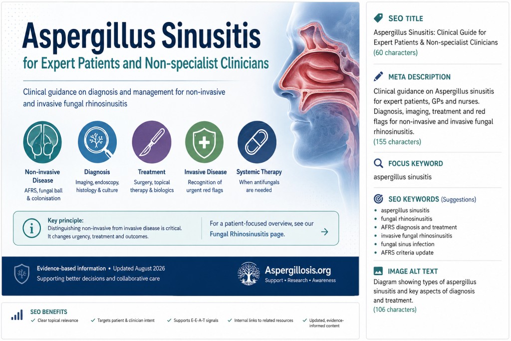
Looking for a general introduction? This article is intended for expert patients, GPs, nurses and other healthcare professionals who want more clinical detail. For a patient-focused overview of the different types of fungal sinus disease, see our Fungal Rhinosinusitis page.
Why fungal sinus disease can be confusing
Aspergillus and other fungi can be associated with several very different diseases of the nose and paranasal sinuses.
The most important distinction is between non-invasive disease, where fungi remain within mucus, crusts or the sinus cavity, and invasive fungal rhinosinusitis, where fungal hyphae grow into sinus tissue.
This distinction matters because the clinical course, urgency and treatment are very different.
| Condition | Typical setting | Tissue invasion? | Usual management principle |
|---|---|---|---|
| Allergic fungal rhinosinusitis (AFRS) | Chronic rhinosinusitis with nasal polyps; often asthma/allergic disease | No | Surgery where required plus long-term control of inflammation |
| Fungal ball | Usually localised to one sinus | No | Endoscopic removal and drainage |
| Saprophytic fungal colonisation | Often crusting or previous sinus surgery | No | Removal of debris and local sinus care |
| Acute invasive fungal rhinosinusitis | Usually major immune suppression or other strong risk factors | Yes | Urgent surgery plus systemic antifungal therapy |
| Chronic invasive fungal rhinosinusitis | More slowly progressive invasive disease | Yes | Surgery plus systemic antifungal therapy |
Allergic fungal rhinosinusitis (AFRS)
Allergic fungal rhinosinusitis is now generally considered a distinct endotype of chronic rhinosinusitis with nasal polyps (CRSwNP).
It is characterised by a strong type 2 inflammatory response, thick eosinophil-rich mucus and non-invasive fungal hyphae within sinus contents.
Although Aspergillus species can be involved, AFRS is not exclusively an Aspergillus disease and other environmental moulds may be identified.
Importantly, AFRS should not be described simply as a fungal infection. The fungus remains outside the tissues; much of the disease results from the inflammatory and immune response associated with its presence.
A 2025 multidisciplinary workshop on AFRS describes the condition as a distinct CRSwNP endotype and highlights major remaining uncertainties around diagnosis, disease mechanisms, recurrence and treatment.
Diagnosis of AFRS
Historically, AFRS has frequently been diagnosed using criteria developed in the 1990s, including the Bent and Kuhn criteria.
Typical features include:
- nasal polyposis
- evidence of fungal sensitisation
- characteristic CT appearances
- eosinophilic or “allergic” mucin
- fungal hyphae within sinus mucus without tissue invasion
However, the diagnostic criteria are increasingly recognised as imperfect. Some require findings obtained only during surgery, some features lack specificity, and patients may not fit neatly into the traditional framework.
The 2025 multidisciplinary workshop concluded that existing diagnostic criteria are outdated and that improved criteria and biomarkers are needed.
A 2025 contemporary review similarly questions the specificity and applicability of the traditional criteria.
In practice, diagnosis therefore depends on the overall pattern of:
- clinical history
- endoscopic findings
- CT imaging
- total IgE and fungal-specific IgE where appropriate
- eosinophil measurements
- allergy testing
- histology and microscopy of sinus contents when available
For more detail, see our 2026 update on changing approaches to AFRS diagnosis.
Imaging in AFRS
CT is the principal imaging investigation for chronic sinus disease. AFRS may produce heterogeneous, dense material within affected sinuses and can sometimes lead to expansion or remodelling of surrounding bone.
Extensive radiological appearances do not necessarily indicate invasive fungal disease. AFRS itself can produce striking sinus expansion and bone changes through chronic inflammation and pressure.
MRI may be useful when disease is extensive or when orbital, skull-base or intracranial involvement needs to be assessed.
Treatment of AFRS
For significant AFRS, endoscopic sinus surgery followed by long-term topical treatment remains the mainstay of management.
Surgery aims to:
- remove thick eosinophilic mucus and fungal debris
- remove obstructing polyps where necessary
- restore sinus ventilation and drainage
- improve access for postoperative saline irrigation and topical corticosteroid treatment
- provide material for histological and microbiological examination
Long-term topical corticosteroids and saline irrigation are important because recurrence is common.
The 2025 contemporary review of AFRS describes surgery followed by topical corticosteroids and saline irrigation as the mainstay of treatment.
Systemic corticosteroids
Systemic corticosteroids may sometimes be used for severe inflammatory disease or recurrence, but their potential benefit has to be balanced against adverse effects, particularly with repeated or prolonged treatment.
They should therefore be regarded as an individual clinical decision rather than an automatic component of treatment for every patient with AFRS.
Antifungal treatment in AFRS
The role of antifungal medicines in AFRS remains uncertain.
This is understandable biologically: AFRS is characterised by non-invasive fungal growth associated with an inflammatory response rather than fungal invasion of tissue.
Older guidance allows consideration of systemic mould-active azoles in refractory or rapidly relapsing disease, but the evidence is limited and antifungal therapy is not routinely required for all patients with AFRS.
Where systemic azoles are used, clinicians need to consider drug interactions, toxicity and therapeutic drug monitoring where appropriate.
Biologic therapy
Biologic medicines targeting type 2 inflammatory pathways are increasingly important in severe asthma and chronic rhinosinusitis with nasal polyps.
There is growing interest in their use for difficult-to-control AFRS, particularly when patients also have severe asthma, recurrent nasal polyposis or other manifestations of type 2 inflammation.
Current AFRS studies are relatively small, so biologics cannot yet be regarded as a replacement for surgery in patients with extensive obstructive disease. Their position in the treatment pathway continues to evolve.
AFRS and the lower airways
Upper and lower airway disease frequently coexist.
Patients with AFRS may also have asthma, and a smaller group may have allergic bronchopulmonary aspergillosis (ABPA).
AFRS and ABPA share several features, including fungal sensitisation, raised IgE and type 2 inflammation, but they affect different anatomical sites:
- AFRS primarily affects the nose and sinuses
- ABPA primarily affects the bronchi and lungs
Persistent upper-airway disease may also make asthma more difficult to control, so considering the patient as having a connected upper and lower respiratory system can be clinically useful.
See our ABPA Knowledge Hub for more information.
Aspergillus fungal ball
An Aspergillus fungal ball is a dense collection of fungal hyphae and debris occupying a sinus without invasion of the surrounding mucosa.
Most are found in the maxillary sinus, although sphenoid and other sinuses may be affected.
The presence of fungal hyphae alone does not establish invasive aspergillosis. Demonstration of tissue invasion is the critical distinction.
Diagnosis
CT may show unilateral sinus opacification containing dense or calcified material. The definitive diagnosis is usually made after removal, with histological examination confirming fungal material without mucosal invasion.
Treatment
Endoscopic surgical removal and adequate sinus drainage are usually sufficient for an uncomplicated fungal ball.
Systemic antifungal treatment is generally not required when there is no evidence of tissue invasion.
The IDSA aspergillosis guideline recommends surgical removal alone for an uncomplicated Aspergillus fungal ball of the paranasal sinus.
Invasive fungal rhinosinusitis
Invasive fungal rhinosinusitis is a fundamentally different disease.
Here, fungal hyphae invade the sinus mucosa and may extend into blood vessels, bone, the orbit, skull base or intracranial structures.
Both Aspergillus and fungi belonging to the Mucorales can cause invasive fungal rhinosinusitis. Identifying the causative organism matters because optimal antifungal therapy differs.
Acute invasive fungal rhinosinusitis
Acute invasive disease usually occurs in people with major predisposing factors such as:
- prolonged or profound neutropenia
- haematological malignancy
- haematopoietic stem-cell transplantation
- solid-organ transplantation
- substantial immunosuppressive treatment
- some severe immunodeficiency states
- poorly controlled diabetes, particularly diabetic ketoacidosis, especially in mucormycosis
The illness may progress rapidly.
Possible warning features include:
- rapidly worsening facial or sinus pain
- facial swelling
- fever
- nasal ulceration or tissue necrosis
- visual disturbance or impaired eye movement
- severe headache
- cranial nerve abnormalities or other neurological symptoms
Suspected acute invasive fungal rhinosinusitis is a medical emergency.
Chronic invasive fungal rhinosinusitis
Chronic invasive fungal rhinosinusitis progresses over a longer period than acute invasive disease but still involves genuine invasion of tissue.
Presentation may include persistent sinus symptoms, facial swelling, proptosis, visual disturbance or evidence of extension beyond the sinus cavity.
The distinction from non-invasive disease cannot safely be made from symptoms alone.
How is invasive disease diagnosed?
Imaging defines the extent of disease, but histopathological evidence of tissue invasion is central to establishing invasive fungal disease.
Assessment may include:
- urgent ENT examination and nasal endoscopy
- CT of the sinuses
- MRI where orbital, skull-base, vascular or intracranial extension is suspected
- biopsy of abnormal tissue
- histopathology with fungal stains
- fungal culture
- molecular identification where available
Culture is valuable because it can identify the organism and sometimes permit susceptibility testing, but a negative culture does not necessarily exclude invasive fungal disease.
Treatment of invasive Aspergillus sinusitis
Management requires urgent multidisciplinary input, commonly involving ENT surgery, infectious diseases or medical mycology, microbiology, radiology and the team managing the patient’s underlying illness.
Treatment usually combines:
- surgical debridement of infected or necrotic tissue where feasible
- systemic antifungal therapy
- management or reversal of underlying risk factors where possible
The IDSA aspergillosis guideline recommends surgery together with systemic antifungal therapy for invasive Aspergillus sinusitis.
Choice of antifungal agent depends on the organism, disease extent, previous antifungal exposure, susceptibility information, organ function, potential drug interactions and individual patient factors.
This is particularly important because invasive fungal rhinosinusitis is not synonymous with invasive aspergillosis: Mucorales require a different therapeutic approach from Aspergillus.
Therapeutic drug monitoring
Several mould-active azole antifungals have variable absorption and metabolism and substantial potential for drug interactions.
When appropriate, therapeutic drug monitoring (TDM) can help establish whether drug exposure is adequate while reducing the risk of toxicity.
TDM should be interpreted alongside the clinical response, organism susceptibility, liver function and interacting medicines rather than as an isolated laboratory result.
Why pathology terminology matters
One of the most important points for non-specialists is that the words “fungus”, “Aspergillus” or “fungal hyphae” in a sinus specimen do not automatically mean invasive aspergillosis.
The key pathological question is whether fungal hyphae are:
- lying within mucus or debris, or
- invading mucosa, blood vessels, bone or other tissue.
This distinction separates conditions such as fungal ball and AFRS from invasive fungal rhinosinusitis and radically changes the treatment required.
When should a non-specialist escalate urgently?
Urgent ENT and specialist assessment should be considered when significant sinus symptoms occur alongside:
- neutropenia or major immune suppression
- haematological malignancy or transplantation
- rapidly progressive facial pain or swelling
- visual symptoms, ophthalmoplegia or proptosis
- black or necrotic nasal tissue
- cranial nerve abnormalities
- severe or unusual headache
- neurological symptoms
- radiological evidence of extension outside the sinus
In this setting, waiting for routine outpatient investigation may be inappropriate.
A useful clinical framework
When fungi are identified or suspected in the sinuses, the first question should not simply be:
“Which antifungal should be used?”
A more useful sequence is:
- What form of fungal rhinosinusitis is present?
- Is there evidence of tissue invasion?
- What organism is involved?
- What underlying host factors are present?
- Is surgery required?
- Is systemic antifungal treatment required?
This prevents non-invasive allergic or saprophytic disease being confused with invasive fungal infection and equally helps ensure that genuinely invasive disease is recognised urgently.
Further information
- Fungal Rhinosinusitis – patient overview
- Allergic fungal rhinosinusitis: how diagnosis is changing
- ABPA Knowledge Hub
References and guidance
- Roland LT, Damask C, Luong AU, et al. Allergic Fungal Rhinosinusitis Diagnosis, Management, Associated Conditions, Pathophysiology, and Future Directions: Summary of a Multidisciplinary Workshop. International Forum of Allergy & Rhinology. 2025;15(6):626–641. PubMed PMID: 40310935.
- Allergic Fungal Rhinosinusitis: A Contemporary Update. 2025. PubMed PMID: 40491076.
- Fokkens WJ, Lund VJ, Hopkins C, et al. European Position Paper on Rhinosinusitis and Nasal Polyps 2020. Rhinology. 2020;58(Suppl S29):1–464. PubMed PMID: 32077450.
- Patterson TF, Thompson GR III, Denning DW, et al. Practice Guidelines for the Diagnosis and Management of Aspergillosis: 2016 Update by the Infectious Diseases Society of America. Clinical Infectious Diseases. 2016;63(4):e1–e60. IDSA guideline.
- Cornely OA, Alastruey-Izquierdo A, Arenz D, et al. Global guideline for the diagnosis and management of mucormycosis. Lancet Infectious Diseases. 2019;19(12):e405–e421. Full text.
Audience: Expert patients, GPs, nurses and non-specialist healthcare professionals
Last reviewed: 17 August 2026
Important: This page provides general information and does not replace assessment or treatment by an ENT, infectious diseases or medical mycology specialist.

Did this page help you?
Tell us in one minute