Nontuberculous Mycobacteria (NTM–MAC) and Aspergillosis
Why these infections sometimes occur together
Audience: Aspergillosis patients, carers, GPs and non-specialist clinicians
Some patients with Allergic Bronchopulmonary Aspergillosis (ABPA) may be investigated for nontuberculous mycobacteria (NTM), because airway damage from ABPA can increase susceptibility to other lung infections.
Key points
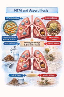
- Nontuberculous mycobacteria (NTM) are environmental bacteria that sometimes infect damaged lungs.
- The most common NTM causing lung disease is the Mycobacterium avium complex (MAC).
- NTM infection and aspergillosis often occur in the same patients because both thrive in damaged airways such as bronchiectasis or lung cavities.
- Some patients with ABPA are investigated for NTM because ABPA can lead to bronchiectasis and impaired mucus clearance.
- NTM infections usually grow very slowly, so treatment is sometimes monitored rather than started immediately.
- Treating NTM and aspergillosis together can be difficult because some NTM antibiotics interfere with antifungal medicines.
- Doctors usually treat the infection causing the most harm first while monitoring the other carefully.
Table of contents
- What are NTM?
- What is Mycobacterium avium complex (MAC)?
- Why NTM infections occur in some people
- What is bronchiectasis?
- Why patients with ABPA may be asked about NTM
- Why NTM and Aspergillus infections often occur together
- The lung infection cycle
- Chronic lung disease as a microbial ecosystem
- Why treatment can be complicated
- When treatment for NTM may be delayed
- How doctors balance treatment decisions
- NTM vs Aspergillosis – comparison table
- Common questions patients ask about NTM and Aspergillus
- When should patients seek medical advice?
- Reducing exposure to NTM in the environment
What are nontuberculous mycobacteria (NTM)?
Nontuberculous mycobacteria (NTM) are bacteria found naturally in the environment.
They live in:
- soil
- water
- dust
- plumbing systems
- shower heads and taps
Unlike tuberculosis, these bacteria are not normally spread between people.
Most people inhale them regularly without becoming ill. However, in some people with damaged lungs, these bacteria can establish a long-term lung infection.
What is Mycobacterium avium complex (MAC)?
The Mycobacterium avium complex (MAC) is the most common cause of NTM lung disease.
This group includes:
- Mycobacterium avium
- Mycobacterium intracellulare
MAC lung disease usually develops slowly over months or years.
Symptoms may include:
- chronic cough
- sputum production
- breathlessness
- fatigue
- weight loss
Because symptoms develop gradually, diagnosis can sometimes take time.
Why NTM infections occur in some people
NTM infections usually develop in people who already have structural lung disease.
Examples include:
- bronchiectasis
- chronic obstructive pulmonary disease (COPD)
- cystic fibrosis
- previous tuberculosis
- severe asthma
- aspergillosis
In these conditions, the lungs have damaged or widened airways, making it harder to clear mucus and microbes.
What is bronchiectasis?
Bronchiectasis is a condition where the airways become permanently widened and distorted.
In healthy lungs, mucus is cleared using:
- mucus movement
- tiny hair-like structures called cilia
- coughing
In bronchiectasis:
- mucus collects in the airways
- microbes become trapped
- infections become more likely
Bronchiectasis is common in patients with Allergic Bronchopulmonary Aspergillosis (ABPA) and other chronic lung diseases.
Why patients with ABPA may be asked about NTM
Some patients with Allergic Bronchopulmonary Aspergillosis (ABPA) are surprised when their doctors start investigating nontuberculous mycobacteria (NTM).
This usually happens because ABPA can lead to bronchiectasis, which increases the risk of other lung infections.
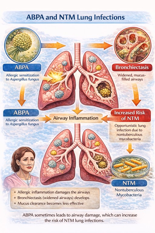
In ABPA:
- inflammation caused by allergic reactions to Aspergillus can damage the airways
- over time the airways may become widened and distorted, causing bronchiectasis
- mucus clearance becomes less effective
When mucus accumulates in the airways, microbes that are normally cleared from the lungs can sometimes persist. These may include:
- nontuberculous mycobacteria (NTM)
- Pseudomonas bacteria
- other organisms that affect bronchiectasis patients
For this reason, doctors sometimes test patients with ABPA for NTM if:
- CT scans show bronchiectasis or nodules
- sputum cultures repeatedly grow unusual organisms
- symptoms worsen without a clear explanation
Importantly, having ABPA does not mean you will develop NTM infection. Most patients with ABPA never develop NTM disease.
However, because the conditions share similar risk factors, doctors sometimes check for both.
Why NTM and Aspergillus infections often occur together
NTM bacteria and Aspergillus fungi both thrive in damaged lungs.
Three factors explain the overlap.
1. Damaged airways trap microbes
When airways are widened or distorted:
- mucus collects
- microbes are not cleared effectively
This allows organisms such as NTM and Aspergillus to persist.
2. Chronic infection causes further lung damage
NTM infection can lead to:
- inflammation
- worsening bronchiectasis
- lung nodules
- sometimes lung cavities
These cavities can then be colonised by Aspergillus, which may lead to chronic pulmonary aspergillosis (CPA).
3. Aspergillus can worsen structural damage
Once Aspergillus becomes established it can cause:
- inflammation
- enlargement of lung cavities
- worsening bronchiectasis
This further damage makes the lungs even more susceptible to infection.
The lung infection cycle
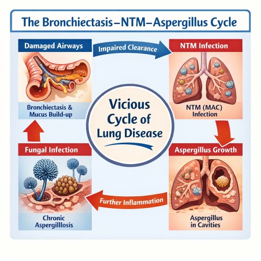
In many patients the relationship between bronchiectasis, NTM and Aspergillus becomes a cycle:
- Lung disease develops
- Bronchiectasis forms
- NTM infection establishes
- Lung damage worsens
- Aspergillus colonises damaged tissue
- Chronic aspergillosis develops
- Lung damage continues
At this stage the lungs may contain multiple organisms simultaneously.
Chronic lung disease as a microbial ecosystem
Doctors increasingly recognise that damaged lungs may contain several interacting microbes rather than a single infection.
Common organisms include:
- Mycobacterium avium complex (MAC)
- Aspergillus species
- Pseudomonas bacteria
- other organisms
For this reason clinicians sometimes describe chronic lung disease as a disturbed lung microbial ecosystem.
Why treatment can be complicated
NTM and aspergillosis treatments can interact.
Typical MAC treatment includes:
- azithromycin or clarithromycin
- ethambutol
- rifampicin
However rifampicin strongly reduces levels of antifungal drugs, including:
- itraconazole
- voriconazole
- posaconazole
These antifungals are commonly used to treat chronic pulmonary aspergillosis.
Because of this interaction, treating both infections at the same time can be challenging.
Other medicines that may interact with rifampicin
Rifampicin affects how the liver processes many medicines. This means it can reduce the effectiveness of several commonly used drugs, including some treatments for heart conditions, blood thinners, hormonal medicines, and certain antidepressants.
Because of this, doctors and pharmacists always review a patient’s medication list before starting rifampicin. Patients should tell their healthcare team about all medicines they take, including over-the-counter medicines, inhalers, and herbal supplements. In most cases, safe alternatives or dose adjustments can be used if needed.
When treatment for NTM may be delayed
Unlike many bacterial infections, MAC often progresses slowly.
Doctors sometimes monitor the infection before starting treatment. This approach is called active monitoring or watchful waiting.
Monitoring may include:
- CT scans
- sputum cultures
- lung function tests
- symptom assessment
Treatment may be delayed if:
- symptoms are mild
- CT scans are stable
- another condition requires more urgent treatment
For example, aspergillosis may be treated first if it is causing the main symptoms or lung damage.
How doctors balance treatment decisions
When both infections are present, clinicians try to identify which infection is currently causing the most harm.
Doctors consider:
Symptoms
- worsening cough
- breathlessness
- fatigue
- weight loss
- haemoptysis (coughing blood)
CT scan findings
- enlarging cavities
- fungal balls
- nodules typical of NTM disease
- worsening bronchiectasis
Laboratory results
- sputum cultures for NTM
- Aspergillus blood tests, such as Aspergillus IgG
If one infection clearly explains the patient’s symptoms, that infection usually becomes the treatment priority.
Treatment plans may then change over time as the balance of disease changes.
NTM vs Aspergillosis – What’s the difference?
| Feature | NTM (MAC) Lung Disease | Aspergillosis |
|---|---|---|
| Type of organism | Bacteria | Fungus |
| Source | Soil, water, plumbing | Airborne fungal spores |
| Spread between people | Rare | Does not spread |
| Typical speed | Slow, chronic infection | Variable |
| Typical CT findings | Nodules, bronchiectasis, cavities | Cavities, fungal balls, airway inflammation |
| Treatment | Long antibiotic courses, often 12–18 months | Antifungal medicines |
| Drug interaction issues | Rifampicin interferes with antifungals | Antifungal levels can be reduced by rifampicin |
Common questions patients ask about NTM and Aspergillus
If MAC grows slowly, why treat it?
Although MAC grows slowly, it can still cause progressive lung damage over time.
Treatment is usually recommended if there is:
- worsening symptoms
- declining lung function
- progressive CT scan changes
Can NTM be present without causing disease?
Yes. Some people have NTM colonisation without active infection.
Doctors diagnose NTM lung disease only when symptoms, imaging findings and repeated cultures all support the diagnosis.
Why do NTM and Aspergillus often occur together?
Both organisms tend to grow in damaged airways, especially where bronchiectasis is present and mucus clearance is poor.
Will both infections always be treated?
Not necessarily. Doctors often treat the infection causing the most immediate problem while monitoring the other.
Does NTM mean my aspergillosis is worsening?
Not necessarily. Both infections occur in damaged lungs, so they may simply share the same environment.
Can NTM lead to aspergillosis?
Sometimes. If NTM infection causes lung cavities or worsening bronchiectasis, these damaged areas may later become colonised by Aspergillus.
Should I worry if my doctor decides not to treat NTM immediately?
Not necessarily. Because MAC often progresses slowly, doctors sometimes choose active monitoring rather than immediate treatment.
When should patients seek medical advice?
People living with aspergillosis, bronchiectasis or NTM infection often have ongoing symptoms such as cough and sputum production. These symptoms may fluctuate and do not always mean the disease is worsening.
However, certain changes should prompt medical review.
Seek medical advice if you notice worsening breathing symptoms
- increasing breathlessness
- a significant increase in cough
- a noticeable increase in sputum production
- sputum becoming thicker, darker or foul-smelling
These symptoms may indicate:
- bacterial infection
- worsening bronchiectasis
- progression of NTM infection
- worsening aspergillosis
Coughing up blood (haemoptysis)
Haemoptysis can occur in both bronchiectasis and aspergillosis.
Seek medical advice if:
- bleeding increases
- blood appears repeatedly
- there is more than a small amount of blood
- bleeding occurs suddenly with breathlessness
Large amounts of blood should be treated as a medical emergency.
Unexplained weight loss or increasing fatigue
Persistent or worsening:
- weight loss
- fatigue
- loss of appetite
may indicate:
- progressive infection
- increasing inflammation
- advancing NTM disease
Fever or feeling unwell
New symptoms such as:
- fever
- chills
- chest discomfort
- feeling generally unwell
may suggest a new infection, such as a bacterial chest infection, which may require treatment.
Rapid change in symptoms
Seek medical advice if you experience:
- sudden worsening breathlessness
- significant chest pain
- new wheezing
- severe fatigue developing quickly
Symptoms that may remain stable
Many people with chronic lung disease experience symptoms that remain relatively stable for long periods, including:
- a chronic cough
- daily sputum production
- mild breathlessness
- intermittent fatigue
Doctors monitor these symptoms over time using:
- CT scans
- sputum cultures
- lung function tests
These investigations help clinicians determine whether infections such as NTM or Aspergillus are stable or progressing.
Reducing exposure to NTM in the environment
Patients with bronchiectasis, ABPA, or other chronic lung diseases sometimes ask whether they should try to avoid environmental exposure to nontuberculous mycobacteria (NTM).
NTM are very common in the natural environment and cannot be completely avoided. They are found in:
- soil and compost
- garden dust
- natural water sources
- tap water and plumbing systems
- showerheads
- hot tubs and spa pools
For most people, the goal is sensible risk reduction rather than strict avoidance. Major lifestyle restrictions are usually not necessary.
Water exposure
NTM can grow in biofilms inside plumbing systems, including showerheads. Small amounts of bacteria may become airborne when water is aerosolised.
Some simple precautions may help reduce exposure:
- avoid frequent use of hot tubs or spa pools
- allow taps or showers to run briefly if they have not been used for several days
- clean showerheads periodically to remove biofilm and limescale
Normal showering and bathing are considered safe for most patients.
NTM infection occurs when bacteria are inhaled into the lungs rather than swallowed. Drinking ordinary tap water is therefore considered safe for most people, and patients are not usually advised to avoid tap water for drinking.
Gardening and soil exposure
NTM bacteria are often present in soil and compost. Gardening can still be enjoyed safely with a few sensible precautions.
- wear gloves when gardening
- avoid inhaling dust from dry compost or soil
- dampen compost before handling to reduce dust
- wash hands after gardening
For people with bronchiectasis or NTM disease, wearing a mask during dusty gardening activities may help reduce inhalation of soil particles.
Reducing dust exposure
Activities that generate dust can increase inhalation of environmental microbes.
Helpful precautions include:
- avoiding sweeping very dusty areas indoors
- ventilating indoor spaces
- wearing a mask during dusty tasks such as handling compost or dry soil
Cleaning showerheads
Cleaning showerheads periodically can help remove limescale and biofilms where microbes may grow.
A simple method is:
- Remove the showerhead if possible.
- Soak it in white vinegar for about 30–60 minutes.
- Gently scrub the spray holes with a small brush.
- Rinse thoroughly.
- Run hot water for 30–60 seconds before use.
If the showerhead cannot be removed, a plastic bag filled with vinegar can be tied around the head so that it soaks.
Cleaning every 1–3 months is usually sufficient.
What is usually not necessary
Experts generally do not recommend major lifestyle changes to avoid NTM exposure. In most cases it is not necessary to:
- avoid showers
- avoid gardening completely
- install specialised water filtration systems
These activities are important for quality of life and general health, and evidence that strict avoidance prevents NTM disease is limited.
The most important protection
For patients with ABPA, bronchiectasis or aspergillosis, the most important protective measures remain:
- good airway clearance
- regular medical monitoring
- prompt treatment of infections
- maintaining overall lung health
Reducing environmental exposure may help slightly, but good management of lung disease remains the most important factor.
Key message
When NTM and Aspergillus infections occur together, treatment decisions focus on which infection is currently causing the most damage, while avoiding harmful drug interactions.
For patients with ABPA, one reason NTM may be discussed is that ABPA can lead to bronchiectasis and impaired mucus clearance, which can make other infections more likely.
Many patients live with these conditions for years with careful monitoring and specialist management.
Author: National Aspergillosis Centre Patient Information Team
Last reviewed: March 2026
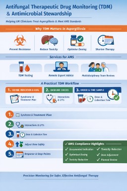
National Aspergillosis Centre, Antifungal Therapeutic Drug Monitoring (TDM), Molecular Resistance Testing & Antimicrobial Stewardship
How the National Aspergillosis Centre Supports UK Clinicians
Long-term antifungal therapy in aspergillosis presents a distinct antimicrobial stewardship (AMS) challenge. Treatment is often prolonged, drug exposure is highly variable, and resistance may emerge during therapy.
The National Aspergillosis Centre (NAC), working closely with the Mycology Reference Centre Manchester (Manchester UK"], provides national expertise through:
-
Therapeutic drug monitoring (TDM)
-
Molecular resistance testing
-
Specialist Advice & Guidance
-
Remote multidisciplinary team (MDT) review
-
Standardised laboratory processes
Together, these services enable UK clinicians to optimise antifungal therapy while aligning with national AMS strategy and antimicrobial resistance (AMR) policy.
The National AMS Framework: Why This Matters
Antifungal stewardship sits within the wider UK antimicrobial resistance strategy.
Key national resources include:
1️⃣ NHS England – Digital Vision for Antimicrobial Stewardship
https://www.england.nhs.uk/long-read/digital-vision-for-antimicrobial-stewardship-in-england/
Emphasises:
-
Data-driven optimisation
-
Decision support
-
Clear documentation
-
Measurable stewardship interventions
2️⃣ Antimicrobial Prescribing & Stewardship Competency Framework
https://www.gov.uk/government/publications/antimicrobial-prescribing-and-stewardship-competencies
Defines clinician responsibilities including:
-
Right drug
-
Right dose
-
Right duration
-
Monitoring for toxicity
-
Review and stop decisions
3️⃣ English Surveillance Programme for Antimicrobial Utilisation and Resistance (ESPAUR)
Supports:
-
National resistance monitoring
-
Stewardship benchmarking
-
Reduction of inappropriate antimicrobial exposure
4️⃣ Chronic Pulmonary Aspergillosis (CPA) Service Specification
https://www.england.nhs.uk/publication/chronic-pulmonary-aspergillosis-service-adults/
This specialised service model explicitly includes:
-
Optimisation of antifungal therapy
-
Toxicity monitoring
-
Therapeutic drug monitoring
Antifungal stewardship is embedded within the commissioned service design.
Why Aspergillosis Requires Enhanced Stewardship
Unlike short-course antibacterial therapy, aspergillosis often involves:
-
Long-term triazole therapy
-
Structural lung disease
-
High interaction burden
-
Emerging environmental resistance
-
Potential for treatment failure despite adequate adherence
Effective stewardship therefore requires both:
-
Assurance of adequate drug exposure (TDM)
-
Assurance of organism susceptibility (molecular testing)
1️⃣ Therapeutic Drug Monitoring (TDM)
Triazole antifungals demonstrate:
-
High pharmacokinetic variability
-
Concentration-dependent toxicity
-
Reduced efficacy if under-dosed
TDM enables:
✔ Early detection of subtherapeutic exposure
✔ Prevention of toxicity
✔ Dose optimisation
✔ Reduction of avoidable escalation
This directly fulfils AMS competency expectations.
2️⃣ Molecular Resistance Testing
Azole resistance in Aspergillus fumigatus is increasingly recognised in the UK.
Through MRCM, NAC supports:
CYP51A Mutation Analysis
Common mutations include:
-
TR34/L98H
-
TR46/Y121F/T289A
These may arise:
-
Environmentally (azole fungicide pressure)
-
During long-term therapy
Phenotypic Susceptibility Testing
Where viable isolates are available:
-
Minimum inhibitory concentration (MIC) testing
-
Clinical interpretation to guide therapy
Why Resistance Testing Is Essential for AMS
If a patient deteriorates despite adequate serum levels:
-
Continuing the same azole is not stewardship
-
Escalating empirically without evidence increases antimicrobial pressure
Molecular confirmation ensures:
✔ Rational switching
✔ Avoidance of ineffective therapy
✔ Contribution to national resistance surveillance
This aligns with ESPAUR and national AMR objectives.
3️⃣ Remote Advice & Guidance & MDT Review
The NAC provides structured national clinician support.
This strengthens stewardship by:
✔ Refining diagnosis
✔ Preventing indication drift
✔ Setting defined review points
✔ Supporting stop decisions
✔ Reducing empirical prolonged therapy
Early specialist review is one of the most effective stewardship interventions.
Integrated Stewardship Model
| Clinical Situation | TDM | Molecular Testing |
|---|---|---|
| Initiation of azole | Yes | Not routine |
| Poor response + low level | Adjust dose | Not primary |
| Poor response + adequate level | Confirm exposure | Essential |
| Long-term therapy | Periodic monitoring | Consider if progression |
| Relapse on therapy | Check level | Strongly consider |
Exposure optimisation + susceptibility confirmation = complete antifungal stewardship.
Practical Workflow for UK Teams
Step 1 – Define Indication
-
Syndrome
-
Treatment objective
-
Planned review date
Step 2 – Baseline Safety Checks
-
Interaction review
-
Liver function tests
-
ECG where appropriate
Step 3 – Perform TDM
Include:
-
Drug
-
Dose
-
Time of last dose
-
Time of sampling
Step 4 – If Clinical Failure Occurs
-
Confirm adequate drug exposure
-
Consider molecular resistance testing
Step 5 – Define Stop/Review Criteria
Avoid open-ended therapy without documented reassessment.
Demonstrating AMS Compliance in Practice
Using NAC-supported services allows Trusts to evidence:
✔ Documented indication
✔ Dose optimisation
✔ Toxicity mitigation
✔ Rational escalation
✔ Defined review intervals
✔ Resistance surveillance contribution
✔ Specialist consultation
This is measurable, defensible antimicrobial stewardship.
Conclusion
Antifungal stewardship in aspergillosis cannot rely on restriction alone.
It requires:
-
Precision dosing
-
Genetic resistance detection
-
Structured specialist review
-
Alignment with national AMS frameworks
Through integrated therapeutic drug monitoring, molecular resistance testing, and national clinical support, the National Aspergillosis Centre provides a UK model for precision antifungal stewardship aligned with national antimicrobial resistance strategy.

🏥 Good News: New AI “Digital Scribe” Helping Doctors Spend More Time With Patients
We’re pleased to share some exciting developments from Manchester University NHS Foundation Trust that could directly improve your experience at clinic appointments.
A new technology called Ambient Voice Technology (AVT) is gradually being introduced across parts of the Trust. Think of it as a secure “digital scribe” that supports your clinician during your consultation.
What does it do?
With your permission, the system listens to the natural conversation between you and your doctor or nurse. It then:
-
Creates the clinical notes automatically
-
Drafts follow-up actions
-
Updates the electronic patient record (*i.e. another reason to use myMFT)
This means your clinician doesn’t need to spend as much time typing or looking at a screen — and can focus more fully on you.
📊 What Have the Early Results Shown?
Colleagues from Manchester University NHS Foundation Trust recently presented results from the Dragon Copilot trial at the Microsoft AI Tour in London.
The findings are encouraging:
✅ 88% of clinicians report saving around 2 minutes per appointment on documentation
✅ 88% say it improves quality and increases face-to-face time with patients
✅ Reduced mental workload for clinicians
✅ Significant reduction in after-clinic administrative work
Two minutes may not sound like much — but across a full clinic list, it adds up. Over time, this could help improve efficiency, reduce waiting times, and improve the overall clinic experience.
💻 How Is It Being Used?
The Dragon Copilot system connects directly into the Trust’s Hive Electronic Patient Record system. It is currently being used in:
-
Outpatient clinics
-
Manchester Royal Infirmary’s Emergency Department
Further expansion is planned in the coming weeks.
❤️ Why This Matters for NAC Patients
For patients with chronic conditions such as aspergillosis, consultations are often detailed and complex. Anything that:
-
Frees up clinician time
-
Improves note accuracy
-
Reduces administrative burden
-
Supports more focused, human interaction
…is a positive step forward.
The aim is not to replace clinicians — but to support them, so your appointment time is spent on what matters most: listening, explaining, planning, and answering your questions.
We’ll continue to keep you updated as this technology develops. It’s encouraging to see innovation being used to strengthen patient-centred care.
If you’d like to learn more, a short video featuring Trust leaders and clinicians was showcased at the Microsoft AI Tour and is available via Trust communications channels.

Watch the World Aspergillosis Day 2026 talks
World Aspergillosis Day 2026 brought together patients, carers, clinicians and researchers to explore how new science and better understanding can improve care for aspergillosis.
Below you can watch the full set of 12 recorded talks from the day, including expert presentations and lived-experience perspectives. You can play them in order, or open the playlist menu to jump to any session.
If you find these videos helpful, please share them — it helps more people living with aspergillosis (and those supporting them) access reliable information and support.
Watch the World Aspergillosis Day 2026 talks
World Aspergillosis Day 2026 brought together patients, carers, clinicians and researchers to explore how new science and better
understanding can improve care for aspergillosis.
Below you can watch the full set of 12 recorded talks from the day, including expert presentations and
lived-experience perspectives. You can play them in order, or open the playlist menu to jump to any session.
If you find these videos helpful, please share them — it helps more people living with aspergillosis (and those supporting them)
access reliable information and support.
Prefer a direct link to the playlist on YouTube?
Open the WAD2026 playlist.

Travelling with Aspergillosis: A Comprehensive Guide to Safe and Stable Travel
This guide is for people living with:
- Chronic Pulmonary Aspergillosis (CPA)
- Allergic Bronchopulmonary Aspergillosis (ABPA)
- Severe asthma (including fungal sensitisation)
- Bronchiectasis
- Fibrotic or structurally abnormal lung disease
Most people with stable disease can travel successfully. The goal is not restriction — it is risk reduction through preparation, environmental awareness, and early action if symptoms change.
Contents
- 1. Understanding Travel Risk in Aspergillosis
- 2. Coordinating With Your Medical Team
- 3. Assessing Stability Before Travel
- 4. Choosing a Destination: Environmental Determinants
- 5. Regional Risk Patterns Explained
- 6. Air Pollution & AQI Monitoring
- 7. Heat, Humidity & Hydration Physiology
- 8. Travel Insurance & Disclosure
- 9. Medication Planning & Contingency Prescriptions
- 10. Specific Considerations for Azole Antifungals
- 11. Air Travel: Physiology & Fit-to-Fly
- 12. Cabin Dryness & Post-Flight Irritation
- 13. Travelling with Oxygen
- 14. Accommodation Risk Reduction
- 15. High-Spore & Dust Exposure Environments
- 16. Infection Prevention
- 17. Haemoptysis Planning
- 18. Red Flag Symptoms
- 19. Advanced Planning Checklist
1. Understanding Travel Risk in Aspergillosis
Travel risk arises from four domains:
- Structural lung vulnerability (cavities, fibrosis, bronchiectasis)
- Inflammatory instability (ABPA activity, asthma control)
- Environmental exposure (humidity, dust, pollution)
- Healthcare accessibility (if deterioration occurs)
Travel is usually safe when disease is stable and exposures are predictable.
2. Coordinating With Your Medical Team
Respiratory Clinic
- Review recent imaging (particularly in CPA)
- Assess haemoptysis history
- Consider fit-to-fly testing if oxygen saturation borderline
- Discuss standby rescue medication
GP
- Ensure medication supply exceeds travel duration
- Provide updated medication summary
- Support vaccination review
- Assist with insurance documentation
3. Assessing Stability Before Travel
Delay travel if within 4–6 weeks of:
- Significant haemoptysis
- Escalating breathlessness
- Recent hospital admission
- New antifungal initiation
Stable inflammatory markers and symptom plateau are reassuring.
4. Choosing a Destination: Environmental Determinants
Key determinants:
- Humidity: promotes indoor mould growth
- Flood history: water damage increases fungal load
- Air pollution: triggers bronchospasm
- Dust burden: irritates inflamed airways
- Healthcare infrastructure: safety net if unwell
5. Regional Risk Patterns Explained
Lower Overall Respiratory Stress
- Scandinavia
- New Zealand
- Canada (outside wildfire season)
Cooler climates limit mould growth; strong building codes reduce damp housing.
Moderate Risk
- Mediterranean Europe
Generally safe when stable; monitor wildfire smoke and heat stress.
Higher Respiratory Stress
- Tropical monsoon climates
- Flood-prone regions
- Highly polluted megacities
- Dust storm zones
Humidity increases fungal proliferation; particulate pollution worsens airway inflammation.
6. Air Pollution & AQI Monitoring
Air pollution can exacerbate cough, bronchospasm, breathlessness and fatigue in people with chronic lung disease. In some urban environments, pollution may pose a greater day-to-day risk than fungal exposure.
The most widely used measure of air quality is the Air Quality Index (AQI), which combines several pollutants into a single score.
Key Pollutants That Matter in Lung Disease
- PM2.5 – fine particulate matter small enough to penetrate deep into the lungs
- PM10 – larger inhalable particles
- Ozone (O₃) – irritates airways, especially in heat
- Nitrogen dioxide (NO₂) – associated with traffic pollution
PM2.5 is particularly important in aspergillosis and severe asthma because it can:
- Trigger airway inflammation
- Increase mucus production
- Worsen bronchospasm
- Reduce exercise tolerance
Reliable Air Quality Monitoring Resources
These sites provide real-time data and forecasts:
- World Air Quality Index (WAQI)
https://waqi.info
Interactive global map with live AQI data for cities worldwide. - IQAir (AirVisual)
https://www.iqair.com
Detailed pollutant breakdowns, 7-day forecasts and wildfire smoke tracking. - UK Daily Air Quality Index (DEFRA)
https://uk-air.defra.gov.uk
Official UK monitoring network with health advice bands.
These platforms also offer mobile apps, which are useful for checking conditions while travelling.
How to Interpret AQI in Practical Terms
| AQI | Category | Practical Advice for Lung Conditions |
|---|---|---|
| 0–50 | Good | Ideal conditions for outdoor activity |
| 51–100 | Moderate | Usually safe; monitor symptoms |
| 101–150 | Unhealthy for sensitive groups | Reduce strenuous outdoor activity; consider indoor plans |
| 151–200 | Unhealthy | Limit time outdoors; avoid exertion |
| 200+ | Very Unhealthy/Hazardous | Stay indoors with filtered air if possible |
For many patients with CPA, ABPA or severe asthma, an AQI above 100 warrants caution. Above 150, limiting outdoor exposure is advisable.
Wildfire Smoke
Wildfire smoke contains high concentrations of PM2.5 and organic particulates. Even patients who are stable at home may experience:
- Increased cough
- Chest tightness
- Increased sputum production
- Fatigue
If travelling during wildfire season:
- Check AQI daily
- Plan indoor activities when levels are elevated
- Use air-conditioned or filtered indoor environments
- Carry rescue inhalers
Urban Pollution vs Rural Dust
Urban areas are more affected by traffic-related pollutants (NO₂, PM2.5), while rural or desert areas may present dust exposure. Both can aggravate inflamed airways.
The risk is cumulative. Short exposure is usually tolerated; prolonged high-level exposure increases the likelihood of symptom flare.
Key principle: checking AQI before and during travel is one of the simplest and most effective risk-reduction steps for people with chronic lung disease.
7. Heat, Humidity & Hydration Physiology
Hot climates place additional physiological stress on people with chronic lung disease.
Why Heat Matters
In warm environments, the body increases sweating and respiratory water loss to regulate temperature. This leads to:
- Increased insensible fluid loss (fluid lost through breathing and skin)
- Reduced plasma volume if intake is inadequate
- Thickening of airway secretions
In bronchiectasis and chronic pulmonary aspergillosis (CPA), mucus clearance is already impaired. Dehydration increases mucus viscosity, making sputum:
- Harder to expectorate
- More likely to stagnate in damaged airways
- Potentially more prone to secondary infection
Patients may notice thicker sputum, increased cough, or chest tightness in hot weather.
Humidity: Helpful or Harmful?
Humidity has mixed effects:
- Moderate humidity can help prevent airway drying.
- High humidity can increase environmental mould growth, particularly indoors if ventilation is poor.
In tropical or monsoon climates, poorly ventilated buildings may have higher fungal spore burdens due to damp conditions.
Heat, Fatigue & Breathlessness
Heat increases cardiovascular demand. The heart works harder to dissipate heat, which can:
- Increase perceived breathlessness
- Increase fatigue
- Reduce exercise tolerance
This does not necessarily indicate worsening lung disease — but it can feel similar.
Hydration Strategy
Practical recommendations:
- Begin hydrating the day before travel
- Drink fluids regularly rather than waiting for thirst
- Aim for pale straw-coloured urine
- Increase intake during flights and hot excursions
Limit:
- Excess alcohol (diuretic effect)
- High caffeine intake
Additional Practical Measures
- Plan outdoor activity early morning or evening
- Rest during peak heat (midday)
- Use air-conditioned environments when available
- Continue airway clearance routines while travelling
Key principle: in chronic lung disease, hydration supports mucus clearance and reduces avoidable exacerbation risk during hot weather.
8. Travel Insurance & Full Medical Disclosure
Travel insurance is not a formality — it is a critical safety net for people with chronic lung disease.
When purchasing insurance, you must declare all pre-existing medical conditions. This typically includes:
- Chronic Pulmonary Aspergillosis (CPA)
- Allergic Bronchopulmonary Aspergillosis (ABPA)
- Severe asthma
- Bronchiectasis
- Pulmonary fibrosis
- Long-term steroid therapy
- Adrenal insufficiency (if present)
- Oxygen use (even if only occasional)
Why Full Disclosure Matters
If you fail to declare a relevant condition, the insurer may:
- Refuse to cover medical treatment abroad
- Decline repatriation costs
- Refuse to reimburse cancelled flights or accommodation
- Invalidate the entire policy
This applies even if the emergency appears unrelated. Insurers may review your full medical history during a claim.
What Insurers Typically Ask
You may be asked:
- Have you been hospitalised in the past 12 months?
- Have you had medication changes recently?
- Have you had haemoptysis?
- Are you awaiting tests or investigations?
- Are you on long-term steroids?
Answer these questions carefully and honestly.
Policies and Stability
Some insurers will decline cover if:
- You have been hospitalised recently
- You are awaiting investigations
- Your condition is considered unstable
This is another reason to travel during a period of clinical stability.
European Travel (UK Patients)
If travelling within Europe, ensure you carry:
- Your GHIC (Global Health Insurance Card)
However, GHIC does not replace travel insurance. It may not cover:
- Private healthcare
- Mountain rescue
- Repatriation to the UK
Practical Tips
- Purchase insurance as soon as you book travel
- Keep written confirmation of declared conditions
- Carry the insurer’s emergency contact number with you
- Inform the insurer early if you require hospital care abroad
In summary: full disclosure protects you. Insurance is only effective if the insurer understands your medical background from the outset.
9. Medication Planning & Contingency Prescriptions
- Carry 1–2 weeks extra supply
- Bring medications in original packaging
- Carry clinic letter
- Consider written rescue plan
10. Specific Considerations for Azole Antifungals
Azoles have significant drug–drug interactions.
- Inform any clinician abroad you are taking an azole
- Avoid grapefruit
- Be aware of sun sensitivity (voriconazole)
- Take itraconazole with food
11. Air Travel: What Actually Happens in the Cabin?
Commercial aircraft cabins are pressurised to simulate an altitude of approximately 6,000–8,000 feet (1,800–2,400 metres).
This means the partial pressure of oxygen is lower than at sea level. For healthy individuals this causes only a small drop in oxygen saturation (typically 3–4%).
Are Most People with Aspergillosis OK to Fly?
Yes — most stable patients fly without difficulty.
People who are:
- Clinically stable
- Not oxygen-dependent
- Without recent haemoptysis
- With resting oxygen saturations ≥95%
generally tolerate commercial flights well, including medium and long-haul travel.
Many patients report anxiety before their first flight after diagnosis, but in stable disease, significant problems are uncommon.
Who Should Consider Fit-to-Fly Testing?
Assessment may be appropriate if you have:
- Resting oxygen saturation consistently below 95%
- Advanced pulmonary fibrosis
- Extensive cavitation
- Significant breathlessness at minimal exertion
- Recent clinical deterioration
The test commonly used is a Hypoxic Challenge Test (HCT), which simulates cabin oxygen conditions to determine whether supplemental oxygen is required during flight.
Where would I have a Hypoxic Challenge Test (HCT)?
In the UK, a Hypoxic Challenge Test is usually arranged through a hospital respiratory physiology department.
You cannot book this test directly. It must be requested by:
- Your respiratory consultant or clinic, or
- Occasionally your GP (who would refer you to a hospital service)
The test is typically performed in:
- A hospital lung function laboratory
- A respiratory physiology unit
- A specialist respiratory centre
During the test, you breathe a gas mixture containing a lower oxygen concentration (usually around 15%) to simulate aircraft cabin conditions. Your oxygen saturation is monitored continuously. If levels fall below safe thresholds, in-flight oxygen may be recommended.
Do Most People Need This Test?
No. Many stable patients with normal resting oxygen saturation (typically ≥95%) do not require hypoxic challenge testing.
The test is generally considered if you:
- Have resting oxygen saturation below 95%
- Have advanced pulmonary fibrosis
- Are already using oxygen
- Have significant exertional desaturation
If you are unsure, ask your respiratory team whether assessment is appropriate for you.
Symptoms During Flight: What Is Normal?
Mild symptoms that can occur in stable patients include:
- Slight increase in breathlessness on walking the aisle
- Fatigue
- Dry cough (often due to low humidity)
These are usually temporary and not dangerous.
Severe symptoms (marked breathlessness at rest, chest pain, dizziness, confusion) are uncommon and require crew notification.
Anxiety vs Physiological Breathlessness
It is very common for people with chronic lung disease to experience heightened awareness of their breathing during flights. The enclosed environment, reduced cabin pressure and awareness of altitude can all increase anxiety.
Anxiety-related breathlessness typically presents as:
- A sensation of not getting a “satisfying” breath
- Chest tightness without wheeze
- Rapid breathing (hyperventilation)
- Tingling in fingers or lips
- Light-headedness
Hyperventilation lowers carbon dioxide levels in the blood. This can cause dizziness, tingling and a feeling of air hunger — even when oxygen levels are normal.
Physiological hypoxia (true low oxygen levels) is less common in stable patients who have been assessed as fit to fly. When it occurs, it is more likely in those with advanced fibrosis, low baseline oxygen saturations, or recent instability.
Features more suggestive of physiological compromise include:
- Persistent breathlessness at rest
- Worsening cyanosis (bluish lips or fingers)
- Marked fatigue or confusion
- Objective low oxygen saturation if measured
For patients who have undergone fit-to-fly assessment and been cleared to travel, significant in-flight hypoxia is uncommon.
Practical Strategies
- Use slow, paced breathing (e.g. inhale for 4 seconds, exhale for 6 seconds)
- Focus on extended exhalation to reduce hyperventilation
- Keep shoulders relaxed and posture upright
- Avoid repeatedly “checking” your breathing
- Remind yourself that mild symptoms are common and expected
Understanding the difference between anxiety-related breathlessness and true hypoxia can significantly reduce distress during flight.
Deep Vein Thrombosis (DVT) Risk
Chronic lung disease does not automatically increase DVT risk, but long-haul immobility does.
General advice:
- Move legs regularly
- Stay hydrated
- Avoid excess alcohol
12. Cabin Dryness & Post-Flight Airway Irritation
Cabin humidity is typically 10–20% (normal indoor comfort is 40–60%).
Low humidity can:
- Dry airway lining
- Reduce mucociliary clearance
- Thicken secretions
- Trigger cough or mild bronchospasm
This is often why people feel they have “caught a cold” the day after flying. In most cases, it is airway irritation rather than infection.
How to Reduce Dryness Effects
- Hydrate well before and during flight
- Limit alcohol and caffeine
- Use isotonic saline nasal spray
- Continue preventer inhalers
- Keep rescue inhaler accessible
- Avoid direct overhead air vents blowing onto your face
- Consider mask use — masks increase humidity of inhaled air
Symptoms typically settle within 24–48 hours.
When to Seek Advice After Flying
Seek medical advice if you develop:
- Progressively worsening breathlessness
- Persistent fever
- Significant haemoptysis
- Chest pain
In stable patients, serious in-flight deterioration is uncommon.
12. Cabin Dryness & Post-Flight Irritation
Cabin humidity is 10–20%.
Dry air:
- Reduces mucociliary clearance
- Thickens secretions
- Triggers cough
- Irritates airways
Hydration and saline sprays reduce symptoms. Post-flight irritation commonly lasts 24–48 hours and does not necessarily indicate infection.
13. Travelling with Oxygen
Confirm airline device approval and battery duration. Plan well in advance.
14. Accommodation Risk Reduction
Request:
- Hard flooring
- No damp odour
- No renovation dust
- Pet-free rooms
Chains Often Reported as Allergy-Conscious
- Hyatt
- Hilton
- Marriott
- Scandic
- Premier Inn
Newer business hotels often have better HVAC filtration.
15. High-Spore & Dust Exposure Environments
- Compost handling
- Construction sites
- Flood-damaged buildings
- Agricultural dust
Avoid heavy inhalation exposure.
16. Infection Prevention
- Hand hygiene
- Avoid close contact with visibly unwell individuals
- Maintain vaccination schedule
17. Haemoptysis Planning
If you have a history of haemoptysis:
- Know your previous pattern
- Carry clinic contact details
- Seek urgent care if volume increases significantly
18. Red Flag Symptoms
- Increasing breathlessness
- New or worsening haemoptysis
- Persistent fever
- Severe chest pain
19. Advanced Planning Checklist
- Travel when stable
- Plan with GP and respiratory clinic
- Carry documentation
- Monitor AQI
- Hydrate on flights
- Avoid damp & heavy dust
- Know red flags
With preparation, most people with stable aspergillosis travel safely and successfully.
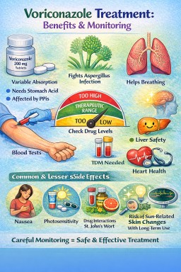
Voriconazole in Aspergillosis
A balanced guide for patients and clinicians
-
Chronic pulmonary aspergillosis (CPA)
-
Allergic bronchopulmonary aspergillosis (ABPA) (selected cases)
-
Invasive aspergillosis
-
Azole-resistant or itraconazole-intolerant cases
It is available orally and intravenously and is often used when a stronger or more reliably absorbed azole is required.
1️⃣ What Voriconazole Does
Voriconazole works by blocking fungal ergosterol synthesis (CYP51 inhibition), which disrupts the fungal cell membrane.
Compared with itraconazole:
-
More potent against Aspergillus
-
More predictable oral absorption
-
More central nervous system penetration
It often produces symptom improvement over weeks, though some effects (e.g. visual symptoms) may occur quickly.
2️⃣ How Long Is Treatment?
In CPA
-
Often 6–12 months or longer
-
Sometimes used as second-line or after intolerance to itraconazole
-
Long-term suppressive therapy may be required
In ABPA
-
Used in selected steroid-dependent or refractory cases
In invasive disease
-
Typically several months depending on response and immune status
3️⃣ Why Blood Level Monitoring Is Essential
Voriconazole has non-linear pharmacokinetics.
Small dose changes can cause large blood level shifts.
Two patients on the same dose may have very different levels due to:
-
Liver metabolism (CYP2C19 genetic variation is important)
-
Drug interactions
-
Age
-
Weight
-
Liver function
If Levels Are Too Low
-
Treatment failure
-
Persistent fungal activity
-
Risk of resistance
If Levels Are Too High
-
Liver toxicity
-
Neurological side effects
-
Visual disturbances
-
Increased interaction risk
Typical Target (Trough)
-
Generally 1–5.5 mg/L (lab dependent)
-
Toxicity risk increases >5–6 mg/L
Levels are usually checked:
-
5–7 days after starting
-
After dose adjustments
-
If side effects occur
-
If clinical response is inadequate
4️⃣ Common Side Effects (Often Mild & Reversible)
Visual Disturbances (Very Common but Usually Harmless)
-
Blurred vision
-
Altered colour perception
-
Light sensitivity
-
“Wavy” vision
These typically:
-
Occur within 30–60 minutes of dosing
-
Last less than an hour
-
Reduce over time
Patients should avoid night driving initially until they understand their response.
Photosensitivity
-
Increased sensitivity to sunlight
-
Sunburn risk
-
Long-term risk of skin damage with prolonged therapy
Sun protection is important.
Gastrointestinal
-
Nausea
-
Abdominal discomfort
5️⃣ Less Common but Important Effects
Neurological
-
Headache
-
Vivid dreams
-
Hallucinations (usually at high levels)
-
Confusion (dose-related)
These are generally reversible with dose adjustment.
Liver Abnormalities
Routine liver function monitoring is required.
Most abnormalities are mild and resolve with dose modification.
Cardiac Effects
Voriconazole can prolong the QT interval.
Caution in patients with:
-
Known arrhythmias
-
Electrolyte imbalance
-
Other QT-prolonging drugs
ECG monitoring may be appropriate in higher-risk patients.
Skin Cancer Risk (Long-Term Use)
With prolonged use (especially >1–2 years):
-
Increased risk of skin squamous cell carcinoma
-
Particularly in transplant recipients
Sun protection and dermatology review are advised for long-term therapy.
6️⃣ Food & Drug Advice
-
Avoid grapefruit
-
Avoid St John’s Wort
-
Take tablets at least 1 hour before or after meals (food reduces absorption)
Voriconazole has many CYP-mediated interactions and requires careful medication review.
7️⃣ Comparison With Itraconazole (Simple Overview)
| Feature | Itraconazole | Voriconazole |
|---|---|---|
| Absorption variability | High | More predictable |
| Visual side effects | Rare | Common but mild |
| Photosensitivity | Rare | More common |
| QT prolongation | Minimal | Possible |
| TDM needed | Yes | Yes (essential) |
Balanced Summary for Patients
Voriconazole is a strong antifungal used when more reliable or potent treatment is needed. Most side effects are manageable and reversible, and blood monitoring keeps treatment safe.
Clinician Checklist
-
Confirm indication and prior azole exposure
-
Check baseline LFTs
-
Review ECG if cardiac risk present
-
Assess drug interactions (CYP2C19, 2C9, 3A4)
-
Arrange trough level at day 5–7
-
Counsel regarding visual symptoms and sun protection
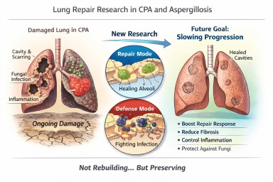
Looking further into the future - could we control lung damage, preserve healthy lung tissue better?
Can Lungs Repair Themselves?
What New Research Means for People with CPA (and Other Aspergillosis)
A recent scientific discovery has helped researchers understand how certain lung cells decide whether to focus on repairing damage or defending against infection. The work, highlighted by the Mayo Clinic and published in Nature Communications, describes a molecular “switch” inside specialised lung cells that influences this balance.
For people living with Chronic Pulmonary Aspergillosis (CPA) — and also those with Allergic Bronchopulmonary Aspergillosis (ABPA) — this kind of research is relevant. But it needs careful explanation.
This is not about rebuilding destroyed lungs.
It is about understanding how to better protect and preserve the lung tissue that remains.
The Discovery: A “Repair vs Defence” Switch
Researchers identified a regulatory circuit in alveolar type II (AT2) cells — specialised cells that:
-
Produce surfactant (which keeps air sacs open)
-
Act as a reserve “repair” population in the lung
-
Can regenerate other essential lung cells after injury
The study showed that these cells operate under tight control. When infection is present, they prioritise defence. When injury needs healing, they can switch into repair mode.
The key insight is that this switch is biologically regulated. It is not random. That means, in theory, it may one day be possible to influence it.
What “Repair” Means — and What It Does Not Mean
When we talk about lung repair in this context, we must be very clear.
It does not mean:
-
Lung cavities caused by CPA will close in the foreseeable future
-
Established fibrosis will melt away
-
Bronchiectasis will reverse
-
Severely distorted lung architecture will rebuild
CPA cavities represent major structural remodelling — destruction of alveoli, scarring, altered blood supply, and thickened pleura. Reconstructing that complex architecture is biologically extremely challenging and not currently realistic within the next decade.
What repair does realistically mean
In chronic lung disease, “repair” is more likely to mean:
-
Supporting survival of remaining alveoli
-
Preventing excessive fibrotic signalling
-
Helping lung lining cells recover more efficiently after inflammation
-
Reducing cumulative injury from repeated infection
-
Slowing progression of structural change
In other words:
Not rebuilding what is gone — but better protecting what remains.
For many people with CPA, this is a crucial distinction.
Why Preservation Is a Major Goal in CPA
CPA usually develops in lungs already weakened by conditions such as tuberculosis, non-tuberculous mycobacteria, chronic obstructive pulmonary disease, or severe pneumonia.
Over time, CPA can lead to:
-
Expanding cavities
-
Progressive scarring
-
Reduced gas exchange
-
Reduced exercise tolerance
Many patients have limited lung reserve. Even small additional losses of functioning lung tissue can significantly increase breathlessness or fatigue.
If future therapies could slow the rate of progression — even modestly — that would meaningfully affect long-term outcomes.
Flattening the decline curve is not trivial. It changes quality of life.
Why This Also Matters in ABPA
In ABPA, repeated inflammatory episodes can lead to:
-
Airway remodelling
-
Mucus plugging
-
Development or progression of bronchiectasis
Better control of inflammatory signalling — combined with improved epithelial recovery — could reduce long-term airway damage.
Again, this is about preservation rather than reversal.
Where Development Has Reached
The current research is still laboratory-based. It used advanced techniques such as:
-
Single-cell sequencing
-
Imaging of lung tissue
-
Preclinical models of injury
No human treatments based on this discovery are yet available.
However, the significance lies in identifying:
-
A defined molecular pathway
-
A controllable regulatory mechanism
-
A clearer understanding of why repair fails in chronic inflammation
That foundational knowledge is what eventually allows targeted drug development.
The Balance Challenge in Aspergillosis
There is an additional complexity in fungal lung disease.
Any attempt to promote repair must not weaken antifungal defence.
The immune system must:
-
Control Aspergillus
-
Avoid causing excessive inflammatory damage
Future therapies would need to strike that balance carefully.
What This Means for Patients Now
This discovery does not change current treatment.
The most effective preservation strategies today remain:
-
Consistent antifungal therapy when indicated
-
Careful inflammatory control
-
Biologic therapies where appropriate
-
Airway clearance
-
Vaccination and infection prevention
-
Avoiding damp and mould exposure
-
Pulmonary rehabilitation
These measures are already forms of lung preservation.
A Realistic and Hopeful Perspective
It is unlikely that cavities from CPA will be repaired in the near future.
It is realistic that within the next 5–10 years we may see improved strategies aimed at:
-
Slowing structural progression
-
Supporting endogenous repair cells
-
Reducing fibrotic signalling
-
Improving recovery after exacerbations
For people living long-term with CPA or ABPA, even incremental preservation could significantly affect independence and quality of life.
The science is still early — but understanding how the lung decides to repair itself is an important step forward.
Reference
Sawhney, A.S., Deskin, B.J., Cai, J. et al. A molecular circuit regulates fate plasticity in emerging and adult AT2 cells. Nat Commun 16, 8924 (2025). https://doi.org/10.1038/s41467-025-64224-1
🧬 How Biologics Are Reshaping Our Understanding of ABPA Subtypes
For many years, Allergic Bronchopulmonary Aspergillosis (ABPA) was viewed as a single condition:
An allergic reaction to Aspergillus fumigatus in the lungs, treated primarily with steroids and sometimes antifungal medication.
Biologic therapies are changing that picture.
They are not just new treatments — they are helping us understand that ABPA may not be one uniform disease, but a spectrum of related inflammatory patterns.
🧠 The Traditional View of ABPA
Historically, ABPA has been defined by:
-
Asthma (or cystic fibrosis)
-
High total IgE
-
Sensitisation to Aspergillus
-
Raised eosinophils
-
Characteristic CT changes (e.g. bronchiectasis, mucus plugging)
The dominant biological explanation was:
A Type 2 (allergic) immune overreaction driven by eosinophils and IgE.
Steroids were used to suppress this immune response.
This model assumed that most patients had broadly similar immune drivers.
💊 What Are Biologics?
Biologics are targeted antibody therapies designed to block specific immune pathways.
In asthma and ABPA, the main targets are:
-
IL-5 (drives eosinophils)
-
IL-5 receptor
-
IL-4 / IL-13 (drive allergic inflammation)
-
IgE
Examples include:
-
Anti–IL-5 therapies (e.g. mepolizumab, benralizumab)
-
Anti–IL-4/IL-13 therapy (e.g. dupilumab)
-
Anti-IgE therapy (e.g. omalizumab)
Instead of broadly suppressing immunity like steroids, they selectively block parts of the allergic pathway.
🔍 What Biologics Are Teaching Us
As biologics have been used in ABPA (often off-label or in specialist centres), an interesting pattern has emerged:
Not all ABPA behaves the same way.
Some patients respond dramatically to anti–IL-5 therapy.
Others respond better to anti–IL-4/IL-13 therapy.
Some show strong IgE-driven disease.
Others appear more mucus-dominant.
This suggests that ABPA may include different inflammatory endotypes (biological subtypes), even if outward symptoms look similar.
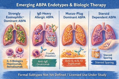
🧩 Possible Emerging ABPA Subtypes
While research is ongoing, clinicians are beginning to recognise patterns such as:
1️⃣ Strongly Eosinophilic-Dominant ABPA
-
Very high eosinophils
-
Frequent exacerbations
-
Often responds well to IL-5 blockade
2️⃣ IgE-Heavy Allergic ABPA
-
Extremely high total IgE
-
Prominent allergic features
-
May respond to anti-IgE therapy
3️⃣ Mucus-Plug Dominant ABPA
-
Recurrent thick mucus impaction
-
Radiological plugging
-
May involve additional inflammatory drivers
4️⃣ Steroid-Dependent ABPA
-
Relapses when steroids reduced
-
Biologics may allow steroid-sparing strategies
These patterns are not yet formal categories, but biologics are revealing that ABPA is biologically more complex than once thought.
🧪 Blood Eosinophils vs Airway Inflammation
Biologics have also highlighted another key insight:
Blood eosinophil levels do not always perfectly reflect what is happening in the lungs.
Some patients:
-
Have modest blood eosinophils
-
But still show eosinophilic airway activity
Biologic response patterns are helping refine how we interpret these markers.
🧠 Moving From “Diagnosis” to “Endotype”
Traditionally, medicine focused on:
Diagnosis (ABPA vs not ABPA)
Biologics are pushing us toward:
Endotype (which immune pathway is dominant in this patient?)
This matters because targeted therapy works best when matched to the dominant pathway.
In future, ABPA may be classified not just by clinical features, but by molecular drivers.
🫁 What This Means for Patients
Biologics offer:
-
Reduced steroid dependence
-
Fewer exacerbations
-
Improved lung function in selected patients
-
Potential improvement in mucus burden
But they also help answer deeper questions:
-
Why do some patients relapse frequently?
-
Why do some have extreme eosinophilia?
-
Why do others have more mucus plugging than inflammation?
They are helping personalise ABPA care.
⚖ Important Caveats
-
Biologics are not currently licensed specifically for ABPA in many countries.
-
Evidence is growing but still developing.
-
They are usually considered in specialist centres.
-
They are not appropriate for every patient.
Steroids and antifungals remain core treatments.
🔭 The Future
Over the next decade, we may see:
-
Better classification of ABPA subtypes
-
Biomarker-guided treatment selection
-
Reduced long-term steroid exposure
-
Improved understanding of mucus plug biology
-
Trials specifically designed for ABPA (rather than extrapolated from asthma)
Biologics are not just new drugs.
They are acting as scientific tools that are reshaping how we think about ABPA itself.
🧠 Key Takeaway
ABPA is no longer seen as one single uniform allergic condition.
Biologic therapies are revealing that:
ABPA is likely a spectrum of related inflammatory patterns — and treatment may increasingly be tailored to the dominant pathway in each individual.
References
Agarwal R, Sehgal IS, Muthu V, Denning DW, Chakrabarti A, Soundappan K, Garg M, Rudramurthy SM, Dhooria S, Armstrong-James D, Asano K, Gangneux JP, Chotirmall SH, Salzer HJF, Chalmers JD, Godet C, Joest M, Page I, Nair P, Arjun P, Dhar R, Jat KR, Joe G, Krishnaswamy UM, Mathew JL, Maturu VN, Mohan A, Nath A, Patel D, Savio J, Saxena P, Soman R, Thangakunam B, Baxter CG, Bongomin F, Calhoun WJ, Cornely OA, Douglass JA, Kosmidis C, Meis JF, Moss R, Pasqualotto AC, Seidel D, Sprute R, Prasad KT, Aggarwal AN. Revised ISHAM-ABPA working group clinical practice guidelines for diagnosing, classifying and treating allergic bronchopulmonary aspergillosis/mycoses. Eur Respir J. 2024 Apr 4;63(4):2400061. doi: 10.1183/13993003.00061-2024. PMID: 38423624; PMCID: PMC10991853.