🧬 How Biologics Are Reshaping Our Understanding of ABPA Subtypes
For many years, Allergic Bronchopulmonary Aspergillosis (ABPA) was viewed as a single condition:
An allergic reaction to Aspergillus fumigatus in the lungs, treated primarily with steroids and sometimes antifungal medication.
Biologic therapies are changing that picture.
They are not just new treatments — they are helping us understand that ABPA may not be one uniform disease, but a spectrum of related inflammatory patterns.
🧠 The Traditional View of ABPA
Historically, ABPA has been defined by:
-
Asthma (or cystic fibrosis)
-
High total IgE
-
Sensitisation to Aspergillus
-
Raised eosinophils
-
Characteristic CT changes (e.g. bronchiectasis, mucus plugging)
The dominant biological explanation was:
A Type 2 (allergic) immune overreaction driven by eosinophils and IgE.
Steroids were used to suppress this immune response.
This model assumed that most patients had broadly similar immune drivers.
💊 What Are Biologics?
Biologics are targeted antibody therapies designed to block specific immune pathways.
In asthma and ABPA, the main targets are:
-
IL-5 (drives eosinophils)
-
IL-5 receptor
-
IL-4 / IL-13 (drive allergic inflammation)
-
IgE
Examples include:
-
Anti–IL-5 therapies (e.g. mepolizumab, benralizumab)
-
Anti–IL-4/IL-13 therapy (e.g. dupilumab)
-
Anti-IgE therapy (e.g. omalizumab)
Instead of broadly suppressing immunity like steroids, they selectively block parts of the allergic pathway.
🔍 What Biologics Are Teaching Us
As biologics have been used in ABPA (often off-label or in specialist centres), an interesting pattern has emerged:
Not all ABPA behaves the same way.
Some patients respond dramatically to anti–IL-5 therapy.
Others respond better to anti–IL-4/IL-13 therapy.
Some show strong IgE-driven disease.
Others appear more mucus-dominant.
This suggests that ABPA may include different inflammatory endotypes (biological subtypes), even if outward symptoms look similar.
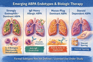
🧩 Possible Emerging ABPA Subtypes
While research is ongoing, clinicians are beginning to recognise patterns such as:
1️⃣ Strongly Eosinophilic-Dominant ABPA
-
Very high eosinophils
-
Frequent exacerbations
-
Often responds well to IL-5 blockade
2️⃣ IgE-Heavy Allergic ABPA
-
Extremely high total IgE
-
Prominent allergic features
-
May respond to anti-IgE therapy
3️⃣ Mucus-Plug Dominant ABPA
-
Recurrent thick mucus impaction
-
Radiological plugging
-
May involve additional inflammatory drivers
4️⃣ Steroid-Dependent ABPA
-
Relapses when steroids reduced
-
Biologics may allow steroid-sparing strategies
These patterns are not yet formal categories, but biologics are revealing that ABPA is biologically more complex than once thought.
🧪 Blood Eosinophils vs Airway Inflammation
Biologics have also highlighted another key insight:
Blood eosinophil levels do not always perfectly reflect what is happening in the lungs.
Some patients:
-
Have modest blood eosinophils
-
But still show eosinophilic airway activity
Biologic response patterns are helping refine how we interpret these markers.
🧠 Moving From “Diagnosis” to “Endotype”
Traditionally, medicine focused on:
Diagnosis (ABPA vs not ABPA)
Biologics are pushing us toward:
Endotype (which immune pathway is dominant in this patient?)
This matters because targeted therapy works best when matched to the dominant pathway.
In future, ABPA may be classified not just by clinical features, but by molecular drivers.
🫁 What This Means for Patients
Biologics offer:
-
Reduced steroid dependence
-
Fewer exacerbations
-
Improved lung function in selected patients
-
Potential improvement in mucus burden
But they also help answer deeper questions:
-
Why do some patients relapse frequently?
-
Why do some have extreme eosinophilia?
-
Why do others have more mucus plugging than inflammation?
They are helping personalise ABPA care.
⚖ Important Caveats
-
Biologics are not currently licensed specifically for ABPA in many countries.
-
Evidence is growing but still developing.
-
They are usually considered in specialist centres.
-
They are not appropriate for every patient.
Steroids and antifungals remain core treatments.
🔭 The Future
Over the next decade, we may see:
-
Better classification of ABPA subtypes
-
Biomarker-guided treatment selection
-
Reduced long-term steroid exposure
-
Improved understanding of mucus plug biology
-
Trials specifically designed for ABPA (rather than extrapolated from asthma)
Biologics are not just new drugs.
They are acting as scientific tools that are reshaping how we think about ABPA itself.
🧠 Key Takeaway
ABPA is no longer seen as one single uniform allergic condition.
Biologic therapies are revealing that:
ABPA is likely a spectrum of related inflammatory patterns — and treatment may increasingly be tailored to the dominant pathway in each individual.
References
Agarwal R, Sehgal IS, Muthu V, Denning DW, Chakrabarti A, Soundappan K, Garg M, Rudramurthy SM, Dhooria S, Armstrong-James D, Asano K, Gangneux JP, Chotirmall SH, Salzer HJF, Chalmers JD, Godet C, Joest M, Page I, Nair P, Arjun P, Dhar R, Jat KR, Joe G, Krishnaswamy UM, Mathew JL, Maturu VN, Mohan A, Nath A, Patel D, Savio J, Saxena P, Soman R, Thangakunam B, Baxter CG, Bongomin F, Calhoun WJ, Cornely OA, Douglass JA, Kosmidis C, Meis JF, Moss R, Pasqualotto AC, Seidel D, Sprute R, Prasad KT, Aggarwal AN. Revised ISHAM-ABPA working group clinical practice guidelines for diagnosing, classifying and treating allergic bronchopulmonary aspergillosis/mycoses. Eur Respir J. 2024 Apr 4;63(4):2400061. doi: 10.1183/13993003.00061-2024. PMID: 38423624; PMCID: PMC10991853.
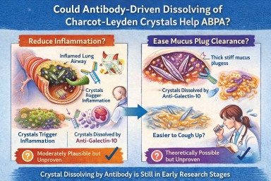
🧬 Could Antibody-Driven Dissolving of Charcot–Leyden Crystals Help ABPA?
Researchers have recently discovered that Charcot–Leyden crystals (CLCs) — the needle-shaped structures formed from the eosinophil protein galectin-10 — are not just debris.
In laboratory studies, specially designed antibodies can dissolve these crystals.
This has raised two important questions:
-
Could dissolving the crystals reduce airway inflammation?
-
Could dissolving them make mucus plugs easier to clear?
Here is what we currently know.
1️⃣ Could dissolving crystals reduce airway inflammation?
What we know
Laboratory and animal studies have shown:
-
Charcot–Leyden crystals can activate immune cells (especially macrophages).
-
They can stimulate inflammatory pathways (including inflammasome signalling).
-
In mouse models, antibodies targeting galectin-10 dissolved the crystals.
-
When crystals were dissolved, airway inflammation decreased.
This suggests that the crystals themselves may amplify inflammation, rather than simply mark it.
What this means biologically
In ABPA and eosinophilic asthma:
-
Eosinophils release galectin-10.
-
Galectin-10 crystallises.
-
Crystals may trigger further immune activation.
-
That leads to more inflammation → more eosinophils → more crystals.
Dissolving the crystals could theoretically interrupt this feedback loop.
How likely is this to help inflammation in humans?
Moderately plausible, but not yet proven.
The biological mechanism is strong.
The animal data are encouraging.
But no human clinical trials have yet shown reduced inflammation through crystal dissolution.
If developed successfully, this approach could:
-
Reduce airway immune activation
-
Lower exacerbation risk
-
Potentially reduce steroid dependence
But at present, it remains investigational.
2️⃣ Could dissolving crystals make mucus plugs easier to cough up?
This is more speculative — but still biologically reasonable.
Why mucus plugs are so thick in ABPA
ABPA mucus plugs contain:
-
Gel-forming mucins
-
DNA from inflammatory cells
-
Dead cells
-
Fungal fragments
-
Eosinophil proteins
-
Charcot–Leyden crystals
The crystals are:
-
Rigid
-
Needle-shaped
-
Structurally stable
When embedded in mucus, they likely increase:
-
Mechanical stiffness
-
Plug density
-
Resistance to deformation
From a physics perspective:
Removing rigid crystalline structures from a gel should reduce stiffness and improve flow.
Do we have direct evidence?
No.
There are currently:
-
No human studies measuring mucus clearance after crystal dissolution
-
No trials showing improved plug expectoration from crystal-targeting therapy
So while it is plausible that dissolving crystals could soften plugs, this has not yet been demonstrated in patients.
3️⃣ How strong is the overall case?
| Outcome | Evidence strength | Likelihood |
|---|---|---|
| Reduced inflammation | Strong biological rationale + animal data | Moderately promising |
| Easier mucus clearance | Biophysical plausibility only | Possible but unproven |
Inflammation reduction is the more evidence-supported target.
Improved plug clearance is plausible but currently theoretical.
4️⃣ How does this compare to existing treatments?
Current therapies (e.g., anti-IL-5 biologics) reduce eosinophils upstream.
That leads to:
-
Less galectin-10 release
-
Fewer crystals forming
-
Reduced inflammation
-
Often improved mucus plugging
So biologics already indirectly reduce crystal burden.
A crystal-dissolving antibody would act downstream, targeting the structural product directly.
This could theoretically:
-
Accelerate resolution of existing plugs
-
Reduce residual inflammatory signalling
But again, this remains in early research stages.
5️⃣ Practical take-home message
At present:
-
Dissolving Charcot–Leyden crystals reduces inflammation in animal models.
-
It is biologically plausible that this could also soften mucus plugs.
-
There is no human clinical proof yet.
-
No approved therapy currently targets the crystals directly.
The concept is scientifically credible — but still under development.
🔭 The Bigger Picture
ABPA is increasingly understood as a condition driven by:
-
Eosinophils
-
Allergic immune signalling
-
Abnormal mucus biology
-
Structural plug formation
Crystal-targeting therapies may eventually become part of a more precise approach to treating eosinophilic airway disease.
But for now, they remain a promising research direction rather than a clinical option.
🔬 Charcot–Leyden Crystals in ABPA and Asthma
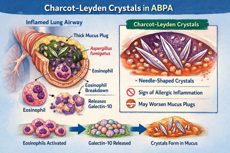
What are they? Why do they form? Do they matter?
If you live with Allergic Bronchopulmonary Aspergillosis (ABPA) or severe asthma, you may see the term Charcot–Leyden crystals in a sputum or pathology report.
They can sound worrying.
They are:
-
Not fungus
-
Not infection
-
Not cancer
They are a sign of a particular type of allergic inflammation in the airways.
🧬 What Are Charcot–Leyden Crystals?
Charcot–Leyden crystals are microscopic, needle-shaped structures found in mucus.
They are made from a protein called galectin-10, which is stored inside a type of white blood cell called an eosinophil.
Eosinophils are immune cells involved in:
-
Allergic asthma
-
ABPA
-
Severe asthma with fungal sensitisation
-
Parasitic infections
When eosinophils are activated and break down, they release galectin-10.
If enough of this protein accumulates in thick airway mucus, it crystallises into visible crystals.
So the crystals are made from your immune cells, not from Aspergillus.
🫁 Why Do They Appear in ABPA?
In ABPA:
-
The immune system overreacts to Aspergillus fumigatus.
-
This triggers a strong allergic (Type 2) immune response.
-
Large numbers of eosinophils move into the airways.
-
Eosinophils break down and release galectin-10.
-
The protein crystallises inside mucus plugs.
The crystals are therefore a footprint of intense allergic inflammation, not fungal invasion.
🌡 Is Most ABPA Eosinophilic?
Yes — almost all classical ABPA is eosinophilic.
ABPA is fundamentally a Type 2 allergic condition, driven by immune pathways involving:
-
IL-4
-
IL-5
-
IL-13
-
IgE
-
Eosinophils
IL-5 in particular stimulates eosinophil production and survival.
Because of this, eosinophils are central to the disease process.
Historically, raised blood eosinophils have been part of diagnostic criteria.
However:
-
Eosinophil counts can fluctuate
-
Steroids can suppress blood levels
-
Eosinophils may still be present in airway mucus even if blood counts appear normal
So ABPA is biologically eosinophilic — even if a single blood test does not show a high count.
True non-eosinophilic ABPA would be unusual and would prompt clinicians to reconsider the diagnosis.
❓ Are Crystals Caused by Aspergillus Infection?
No.
They are caused by the immune reaction to Aspergillus — not by the fungus itself.
They can also be seen in:
-
Severe eosinophilic asthma
-
Parasitic infections
-
Other allergic lung conditions
They reflect eosinophil activity, not fungal growth.
🧠 Why Don’t All People with Asthma Develop These Crystals?
Asthma is not one single disease. It has different inflammatory patterns.
Type 2 (Eosinophilic) Asthma
This involves high eosinophils and allergic pathways.
Common in:
-
Allergic asthma
-
ABPA
-
Severe eosinophilic asthma
These patients can develop Charcot–Leyden crystals.
Non–Type 2 (Non-Eosinophilic) Asthma
This includes:
Neutrophilic asthma
Driven by neutrophils rather than eosinophils.
Paucigranulocytic asthma
Very few inflammatory cells present.
In these forms:
-
Eosinophils are low
-
Galectin-10 is not released in large amounts
-
Crystals are unlikely to form
🧱 Do Charcot–Leyden Crystals Make Mucus Plugs Worse?
Possibly.
Research suggests they may:
-
Increase mucus thickness
-
Contribute mechanically to airway blockage
-
Stimulate further inflammation
For many years they were thought to be harmless debris.
Modern studies suggest they may actively amplify inflammation when present in large amounts.
🎯 Do They Have a Purpose?
Eosinophils evolved mainly to help fight parasitic infections.
Galectin-10 probably has immune signalling roles inside cells.
However, when large amounts are released into thick airway mucus, crystallisation appears to be a by-product of excessive immune activity rather than a useful defence.
In ABPA and allergic asthma, they are more likely part of the problem than part of the solution.
💧 Can Their Formation Be Reduced?
Hydration alone does not stop them forming.
Drinking fluids helps:
-
Keep mucus less sticky
-
Support airway clearance
But it does not prevent eosinophils releasing galectin-10.
What reduces crystal formation?
Reducing eosinophilic inflammation:
-
Corticosteroids
-
Anti-IL-5 biologics
-
Anti-IL-4/IL-13 biologics
When eosinophil numbers fall:
→ Less galectin-10 is released
→ Fewer crystals form
Antifungal treatment in ABPA may indirectly help by reducing allergic stimulation, but the main driver is the immune response.
📊 Do They Change Treatment?
Not directly.
Doctors base treatment on:
-
Symptoms
-
Blood eosinophils
-
Total IgE
-
Imaging
-
Lung function
-
Exacerbation history
Crystals support the diagnosis of eosinophilic inflammation but do not determine treatment alone.
🔎 What Do They Tell Us?
Charcot–Leyden crystals tell us:
-
The airway inflammation is eosinophilic.
-
The immune response is strongly allergic.
-
Mucus plugging risk may be higher.
They are a marker of immune overreaction, not infection severity.
🧠 Key Points to Remember
-
They are made from proteins released by eosinophils.
-
They are not Aspergillus.
-
They do not mean invasive fungal infection.
-
Most classical ABPA is eosinophilic.
-
They are unlikely in non-eosinophilic asthma.
-
Reducing eosinophils reduces their formation.
-
Hydration helps clearance but does not prevent formation.
In simple terms:
Charcot–Leyden crystals are microscopic signs that the immune system is working too hard in the airways.

Invitation: Patient & Carer Discussion on Living with ABPA. New type of treatment.
🕙 10:00am, Thursday 12th
Get details on how to join us by clicking on the link below and choosing Thursday 12th Patients Support Meeting - you will be sent a link to the meeting via email.
https://outlook.office.com/book/[email protected]/
We are inviting people living with Allergic Bronchopulmonary Aspergillosis (ABPA), and those who care for them, to take part in an open, informal online discussion with argenx, a research-focused biotechnology company.
argenx would like to listen directly to patients and carers to better understand what day-to-day life with ABPA is really like. There is no need to prepare anything in advance — you are welcome simply to listen, or to share as much or as little as you feel comfortable.
They are particularly interested in hearing about:
-
Patients’ and carers’ journeys living with ABPA
-
Which symptoms are most burdensome in everyday life (for example breathlessness, cough, fatigue, thick mucus or mucus plugs)
-
Where current treatments fall short from a patient or carer perspective
-
What would make patients or carers feel motivated or reassured about taking part in a future clinical trial of a new ABPA therapy
The purpose of this conversation is to help researchers design future studies that reflect what matters most to patients, including which outcomes are meaningful and how trials can be made more patient-friendly.
📅 Date: Thursday 12th
🕙 Time: 10:00am
💬 Format: Open, informal discussion
📝 Preparation: None required
If you are living with ABPA, or care for someone who is, and would be interested in attending, please let us know.
A short explainer: what is ARGX-118?
argenx is developing an investigational (research-stage) treatment called ARGX-118. It is not yet a licensed medicine and is not currently available outside of research studies.
In ABPA, many people experience very thick, sticky mucus and mucus plugs that block airways and contribute to breathlessness, cough, and flare-ups. Research has shown that this mucus can sometimes contain microscopic crystals formed from proteins released by certain white blood cells involved in allergic inflammation. These crystals can make mucus denser and harder to clear.
ARGX-118 is designed to target and break down these crystals, with the aim of making mucus less thick and easier to clear from the lungs. This is a different approach from current treatments, which mainly focus on suppressing inflammation (such as steroids or biologics) or reducing fungal burden (antifungal medicines).
Because ARGX-118 is still in early development, we do not yet know how effective it will be, who might benefit most, or how it would fit alongside existing treatments. That is exactly why argenx wants to hear from patients and carers now — to understand real-world symptoms, treatment gaps, and what would genuinely matter if a future clinical trial were developed.
👉 Attending this meeting does not commit you to any trial and will not affect your care. It is simply an opportunity to share experiences and help shape future research, if you wish.
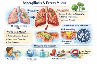
Airways mucus and aspergillosis
A clear, patient-friendly explainer
People living with aspergillosis often say that mucus is one of the hardest symptoms to manage — thick sputum, coughing fits, plugs that feel “stuck”, and flare-ups that seem to come out of nowhere. This explainer brings everything together in one place: what mucus is for, why aspergillosis causes so much of it, why it becomes abnormal, and what current and future treatments aim to do.
1. What is airway mucus and why do we need it?
Mucus is normal, healthy, and essential. Everyone produces it all the time.
Its main roles are to:
-
Trap inhaled particles (dust, spores, bacteria, pollution)
-
Protect the airway lining from drying and irritation
-
Support the immune system
-
Clear the lungs, using tiny moving hairs (cilia) that sweep mucus upwards so it can be swallowed or coughed out
(this clearance system is called the mucociliary escalator)
In healthy lungs:
-
Mucus is thin
-
Produced in small amounts
-
Cleared without you noticing it
2. Why aspergillosis causes excessive mucus
In aspergillosis, the lungs are under ongoing stress. Several factors combine:
Persistent immune activation
The immune system keeps reacting to Aspergillus material in the airways. Even when the fungus is controlled, inflammation can persist.
Allergic-type inflammation (especially in ABPA)
Allergic immune responses strongly stimulate mucus-producing cells, leading to:
-
Large volumes of mucus
-
Very sticky or rubbery sputum
Airway damage
Conditions commonly associated with aspergillosis (such as bronchiectasis or long-standing asthma) cause:
-
Widened or damaged airways
-
Poor mucus clearance
-
Pools of mucus that are hard to shift
Slowed clearance
Inflammation and infection impair cilia, so mucus:
-
Moves more slowly
-
Sits in the lungs longer
-
Becomes thicker and harder to clear
➡️ What starts as a protective response becomes a self-perpetuating problem.
3. Why thick mucus causes symptoms
Excess or abnormal mucus can:
-
Block airways → breathlessness and wheeze
-
Trigger coughing → especially overnight or on waking
-
Trap infection → repeated flare-ups
-
Reduce oxygen exchange
-
Increase fatigue and chest discomfort
Many patients describe it as:
“Glue-like”, “stringy”, “rubbery”, or “impossible to move”
4. Mucus plugs and crystals – why some mucus is so hard to clear
Mucus plugs
When mucus becomes very thick, it can:
-
Form plugs that partially or completely block airways
-
Show up on CT scans
-
Worsen breathlessness suddenly
Charcot–Leyden crystals
In allergic and eosinophilic airway disease (including allergic bronchopulmonary aspergillosis):
-
Breakdown products of allergic immune cells can form microscopic crystals
-
These crystals make mucus:
-
Stiffer
-
More irritating
-
Harder to clear
-
Their presence is a sign of ongoing allergic inflammation, not infection alone.
5. Why managing mucus really matters
Mucus is not just an inconvenience. Poor mucus control can:
-
Increase infection risk
-
Drive repeated exacerbations
-
Worsen lung damage over time
-
Reduce quality of life and sleep
-
Increase hospital admissions
For aspergillosis, mucus management is core treatment, not optional.
6. What helps now (current approaches)
A. Thin the mucus
-
Good hydration
-
Nebulised saline (normal or hypertonic)
-
Selected mucolytic medicines (used carefully)
B. Move it out
-
Regular airway clearance physiotherapy
-
Breathing techniques (e.g. active cycle breathing)
-
Oscillating devices (flutter, Acapella, Aerobika)
-
Gentle, regular physical activity where possible
C. Reduce inflammation
-
Inhaled corticosteroids (when appropriate)
-
Oral steroids (used cautiously)
-
Biologic therapies for selected allergic or eosinophilic disease
-
Antifungal treatment when fungal burden is contributing
D. Treat infections early
-
Bacterial infections thicken mucus further
-
Prompt treatment reduces long-term damage
7. What research is doing differently (emerging therapies)
Research is moving beyond simply “loosening mucus”.
1. Reducing mucus production at source
Scientists are developing drugs that aim to:
-
Switch off excessive mucus secretion
-
Preserve normal protective mucus
This targets the mucus-producing cells directly.
2. Blocking the signals that drive over-production
Inflammation sends chemical signals telling airways to make more mucus. New treatments aim to:
-
Calm allergic and immune pathways
-
Prevent expansion of mucus-producing cells
Some current biologic therapies already reduce mucus indirectly; future drugs will be more precise.
3. Changing mucus structure
Instead of thinning everything, researchers are studying ways to:
-
Loosen the internal “mesh” of mucus
-
Prevent dense plugs from forming
-
Restore normal movement by cilia
4. Targeting mucus crystals
In allergic aspergillosis, research is exploring how to:
-
Reduce crystal formation
-
Calm the specific immune responses that create them
5. New inhaled and physical approaches
Early trials are testing:
-
Inhaled therapies designed to mobilise secretions
-
Treatments that improve airflow behind mucus plugs
6. Precision medicine
Future mucus treatments are likely to be:
-
Personalised
-
Based on inflammation type, fungal involvement, airway damage, and immune markers
Two people with aspergillosis may have very different mucus drivers — and need different solutions.
8. What this means for patients today
-
There is no single “anti-mucus cure” yet
-
Promising therapies are in research and early trials
-
Safety and long-term effects must be proven first
For now:
-
Regular airway clearance remains essential
-
Treating inflammation and infection promptly is crucial
-
Understanding why your mucus behaves as it does helps guide treatment
Key messages to remember
-
Mucus is normally protective
-
Aspergillosis turns a helpful system into a problem
-
Thick, sticky mucus reflects ongoing inflammation and airway damage
-
Crystals signal allergic involvement, not just infection
-
Research is moving toward preventing abnormal mucus formation, not just thinning it
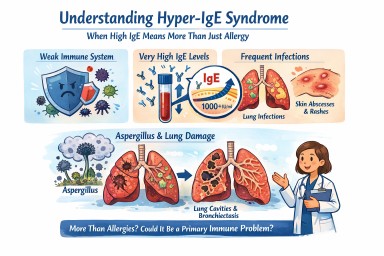
Hyper-IgE syndrome
A patient-friendly guide (and why it matters if you have aspergillosis)
It is not the same as having lots of allergies, even though it can look very similar at first.
What is IgE, and why does it matter?
IgE is usually involved in allergies and asthma.
In Hyper-IgE syndrome:
-
IgE levels are extremely high (often many thousands)
-
But the immune system is unbalanced
-
This makes infections—especially in the lungs and skin—harder to control
So IgE is high, but protection is weak.
How might Hyper-IgE syndrome affect everyday life?
Not everyone has the same symptoms, but common features include:
Lung and chest problems
-
Repeated chest infections (often from a young age)
-
Ongoing cough, breathlessness and mucus
-
Lung damage such as bronchiectasis
-
Lung cavities that can later become infected by moulds such as Aspergillus
Skin and infection problems
-
Long-standing eczema or very sensitive skin
-
Recurrent skin infections or boils
-
Infections that keep coming back or take a long time to clear
Other clues (in some people)
-
Frequent infections in childhood
-
Bone or joint problems
-
Dental issues (for example baby teeth not falling out on time)
Why is this important for people with aspergillosis?
For many people, Aspergillus causes allergy or irritation.
In Hyper-IgE syndrome:
-
The immune system struggles to control moulds
-
Aspergillus can behave more like a true infection, not just an allergy
-
Lung damage can happen more easily and progress faster
This means doctors may need to:
-
Monitor lungs more closely
-
Treat fungal disease earlier and for longer
-
Be cautious with repeated or long-term steroid use
Specialist centres such as the National Aspergillosis Centre are often involved when aspergillosis and immune problems overlap.
Isn’t this just severe allergy or ABPA?
Hyper-IgE syndrome can look similar to:
-
Severe allergic asthma
-
Allergic Bronchopulmonary Aspergillosis (ABPA)
The key difference is that in Hyper-IgE syndrome:
-
The immune system itself is faulty
-
High IgE is part of a wider immune problem
-
Treating allergy alone may not be enough
Some people are treated for asthma or ABPA for years before this possibility is considered.
How is Hyper-IgE syndrome treated?
There is no single cure, but good treatment can make a big difference. The aim is to prevent infections, protect the lungs, and reduce symptoms.
1. Preventing infections (most important)
Because the immune system does not fight germs normally:
-
Some people take regular low-dose antibiotics
-
Others use antibiotics early and promptly when infections start
For people with aspergillosis:
-
Antifungal medicines may be needed
-
Monitoring is usually closer and longer-term
2. Protecting the lungs
Many people develop bronchiectasis or lung damage, so care often includes:
-
Airway clearance physiotherapy
-
Saline nebulisers to help clear mucus
-
Regular sputum tests
-
Early treatment of flare-ups
The goal is to stop the cycle of:
infection → inflammation → permanent lung damage
3. Managing inflammation and allergy (carefully)
People may also have asthma-like symptoms, eczema and multiple allergies.
-
Steroids can help symptoms, but long-term or frequent use can increase infection risk
-
Doctors usually try to keep steroid doses as low as possible
Biologic treatments (such as anti-IgE medicines):
-
May help some people
-
Do not fix the immune problem
-
Are considered on an individual basis, usually in specialist centres
4. Skin care
-
Regular moisturising
-
Prompt treatment of infected eczema
-
Good skin care helps reduce infection risk
How is Hyper-IgE syndrome diagnosed?
Diagnosis usually involves:
-
A detailed review of your medical history (often including childhood infections)
-
Blood tests of immune function
-
Referral to an immunology specialist
-
Sometimes genetic testing
Does having high IgE mean I definitely have this?
No.
Hyper-IgE syndrome is rare.
But it may be worth asking about if:
-
Your IgE has always been extremely high
-
You’ve had repeated infections for many years
-
You have bronchiectasis without a clear cause
-
Aspergillosis seems unusually persistent or severe
-
Standard asthma or allergy treatments don’t fully explain your symptoms
Key message
Very high IgE does not always mean “just allergy.”
In a small number of people, it reflects a deeper immune problem that changes how aspergillosis behaves and how it should be treated.
If your illness doesn’t quite fit the usual labels, it is reasonable to ask whether an immunology review would help.

Sinusitis in Patients with ABPA
When to suspect it, when to investigate, and when to refer
Why this matters
Patients with allergic bronchopulmonary aspergillosis (ABPA) are usually managed as having a lung disease. Diagnosis, monitoring, and treatment focus appropriately on the chest, immunology, and asthma control.
However, ABPA occurs within a single continuous airway, extending from the nose and sinuses to the lungs. Disease in the upper airway can coexist with, exacerbate, or complicate lower airway inflammation — yet sinus disease is not routinely assessed in ABPA care pathways.
This article outlines:
-
What is known about sinus disease in this context
-
Which symptoms should raise suspicion
-
When investigation or ENT referral should be considered
-
What GPs and non-specialists can reasonably do
The united airway: a brief reminder
The upper and lower airways share:
-
Type 2 (eosinophilic) inflammation
-
Immunoglobulin E–mediated immune responses
-
Common triggers, including allergens and fungi
Chronic rhinosinusitis is common in asthma and severe asthma, and treatment of sinus disease can improve lower airway outcomes in some patients.
ABPA sits within this same inflammatory spectrum, even though its management is lung-centred.
Sinus disease in ABPA: what is (and isn’t) known
What we know
-
Chronic rhinosinusitis is common in patients with asthma and severe asthma
-
Sinus disease may be symptomatic or relatively silent
-
ABPA guidelines do not mandate routine ENT review or sinus imaging
-
ENT involvement, therefore, varies widely between centres
What we do not know
-
Whether routine ENT assessment improves ABPA outcomes
-
Which ABPA patients benefit most from sinus intervention
-
The optimal timing for ENT referral in ABPA
As a result, clinical judgement remains central.
Symptoms that should prompt consideration of sinus disease
Sinusitis in ABPA patients does not always present with classic “blocked nose and facial pain”.
Key symptoms include:
Common but often overlooked
-
Persistent post-nasal drip
-
Foul, bitter, metallic, or “infected” taste in the mouth
-
Throat clearing, chronic cough
-
Thick or sticky mucus sensation
-
Symptoms are worse on waking or lying flat
More typical sinonasal features
-
Nasal blockage or congestion
-
Facial pressure or fullness
-
Reduced or altered sense of smell
-
Nasal crusting or discharge
Contextual clues
-
Poor durability of response to steroids or antifungals
-
Recurrent “flares” without clear chest triggers
-
Coexisting severe asthma or nasal polyps
-
Symptoms are worse in damp or mould-affected housing
A persistent foul taste in the mouth is a recognised symptom of chronic sinus disease, usually due to post-nasal drainage of inflamed secretions.
Damp homes and sinus disease
Living in damp or mould-affected environments is associated with:
-
Higher rates of chronic rhinosinusitis
-
Upper airway irritation and inflammation
-
Allergic sensitisation to fungal spores
In most cases, this results in inflammatory or allergic sinusitis, not invasive fungal infection.
Fungal involvement may act as an immune trigger, even when not labelled as “fungal sinusitis”.
Fungal sinusitis: rare vs under-recognised
It is important to distinguish between entities:
| Type | Frequency | Key point |
|---|---|---|
| Invasive fungal sinusitis | Rare | Usually immunocompromised; dramatic presentation |
| Fungal ball (mycetoma) | Uncommon | Usually obvious on CT |
| Allergic fungal rhinosinusitis | Likely under-recognised | Requires active suspicion |
Allergic fungal rhinosinusitis overlaps biologically with ABPA:
-
IgE-mediated
-
Eosinophilic inflammation
-
Thick allergic mucin
It is not routinely sought, so it may be under-diagnosed in at-risk groups.
What GPs and non-specialists can reasonably do
1. Take upper airway symptoms seriously
Especially in ABPA or severe asthma patients with:
-
Persistent post-nasal symptoms
-
Foul taste
-
Recurrent unexplained deterioration
2. Examine the nose and throat
-
Look for polyps, discharge, and crusting
-
Note mouth breathing or altered voice quality
-
Check dentition (to exclude dental causes)
3. Consider imaging when symptoms persist
-
CT sinuses (not plain X-ray) is the imaging of choice
-
Particularly appropriate if symptoms last >8–12 weeks or recur
4. Refer to ENT when:
-
Symptoms are persistent or progressive
-
CT shows significant sinus disease
-
There is a poor response to standard medical therapy
-
There is diagnostic uncertainty
Referral does not imply surgery — ENT input may be diagnostic or medical.
What this article is not saying
-
It does not suggest that all ABPA patients need an ENT referral
-
It does not claim that sinus treatment improves ABPA outcomes
-
It does not override existing guidelines
It does suggest that earlier consideration of the upper airway is reasonable in selected patients.
Key take-home points for clinicians
-
The airway functions as a single inflammatory system
-
Sinus disease may be subtle, under-reported, or atypical
-
A foul taste in the mouth is a meaningful symptom
-
Damp or mould exposure increases sinus disease risk
-
ENT referral is appropriate when symptoms persist or recur
-
Evidence gaps remain — but clinical vigilance is justified
In summary
ABPA is managed as a lung disease, but patients live with a whole airway.
Recognising when sinus disease may be contributing can help explain persistent symptoms and guide appropriate referral — without over-investigation or over-treatment.

ABPA and Work: What a Patient Poll Tells Us About Employment, Health, and Real-World Impact
An article for patients, GPs, and non-specialist healthcare professionals
Allergic bronchopulmonary aspergillosis (ABPA) is often discussed in terms of lung function, immunology, and imaging. Far less often do we talk about its impact on everyday life, particularly on a person’s ability to work.
A poll run within the National Aspergillosis Centre patient community asked a simple but powerful question:
Who is still able to work while living with ABPA – and who has had to stop or retire?
The responses provide an important insight into the functional and socioeconomic burden of ABPA.
Key findings from the poll (patient-reported)
-
Working full time: 17%
-
Working part time (days or hours): 18% combined
-
Not working: 30%
-
Retirement age: 21%
-
Retired early for health reasons: 12%
-
Currently on sick leave / full-time carer / pre-diagnosis: small but notable groups
Even allowing for the informal nature of a social media poll, the overall pattern is clear.
What this tells us
1. Sustained full-time work is uncommon in ABPA
Fewer than one in five respondents were able to work full time. Even among those still working, many described reduced hours, flexible arrangements, or fragile employment dependent on day-to-day health.
ABPA is often incompatible with predictable, high-demand working patterns.
2. ABPA frequently leads to work loss or early retirement
A substantial proportion of respondents were either:
-
No longer working at all, or
-
Retired earlier than planned specifically because of health
This is particularly striking given that ABPA often affects people during their working years and may coexist with asthma, bronchiectasis, or long-term steroid use.
3. “Retirement age” can hide health-forced exit
Some respondents selected “retirement age,” but accompanying comments revealed that many:
-
Left work earlier than expected
-
Changed careers or reduced responsibilities years before retirement
-
Worked through ill health until they no longer could
This matters when interpreting employment statistics: health-driven work loss may be underestimated.
4. Unpaid work and instability are often overlooked
The poll also highlighted:
-
People currently on prolonged sick leave
-
Full-time unpaid carers
-
Individuals still awaiting diagnosis but already struggling to work
These groups are frequently invisible in employment data, yet represent significant personal and societal impact.
Why ABPA affects the ability to work
For patients and non-specialists, it is important to understand that work difficulties in ABPA are not simply due to “asthma symptoms.”
Common contributors include:
-
Chronic breathlessness and cough
-
Severe fatigue and post-exertional exhaustion
-
Recurrent chest infections
-
Steroid side-effects (muscle weakness, bone disease, mood changes, diabetes risk)
-
Unpredictable flare-ups requiring rest, antibiotics, or hospital care
-
Cognitive and emotional burden of long-term illness
Together, these make consistent attendance, physical work, and high cognitive load difficult to sustain.
Implications for patients
-
Difficulty working is not a personal failure
-
Many others with ABPA face similar challenges
-
Adjustments, reduced hours, or stopping work altogether may be medically appropriate
-
Asking for support is reasonable and justified
Implications for GPs and non-specialist clinicians
-
Employment status should be considered a key outcome of disease control
-
Fit notes, occupational health input, and benefits documentation are part of holistic care
-
ABPA is a fluctuating condition – patients may cope for periods and then deteriorate
-
Statements such as “lung function is stable” do not always reflect real-world functioning
Understanding the work impact helps clinicians better support patients in consultations, reports, and advocacy.
Implications for systems and policy
This poll reinforces that ABPA carries a significant socioeconomic burden, including:
-
Reduced workforce participation
-
Early retirement
-
Increased reliance on health and social support systems
Any assessment of disability, employment capability, or long-term planning must take into account:
-
Variability over time
-
Treatment burden
-
Side-effects of necessary medications
In summary
This patient poll sends a consistent message:
ABPA commonly limits the ability to work, often leading to reduced hours, unstable employment, or early exit from the workforce.
For patients, this experience is shared and valid.
For clinicians, it is a reminder that ABPA is not just a radiological or immunological diagnosis, but a life-limiting condition with real-world consequences.

Why do some people cough up long, tube-shaped pieces of mucus?
In several chronic lung conditions, the airways can become inflamed and produce thick mucus.
When this mucus sits in the bronchial tubes, it can sometimes harden into a cast shaped exactly like the airway.
People often describe these casts as:
-
long, ribbon-like or “snakeskin” pieces
-
rubbery or stretchy
-
white, yellow, or green
-
shaped like the inside of a tube
Coughing one up can feel dramatic but is usually a sign that your lungs are finally able to clear a blockage.
What does it mean if a cast has black flecks or dark spots?
This can look alarming, but several common, mostly harmless explanations exist.
1. Old or dried blood
Tiny amounts of bleeding from irritated airways can dry and turn:
red → brown → black
This often appears as tiny black dots or threads.
2. Inhaled particles
Dust, soot, pollution, or smoke can get trapped in mucus deeper in the lungs and show up as dark specks.
3. Debris from infection or inflammation
Long-standing inflammation can cause:
-
darkened mucus fragments
-
tiny bits of fungal, bacterial or biofilm material
-
oxidised (darkened) mucus layers
These often look like pepper-like flecks and are not dangerous on their own.
4. Oxidation or ageing of thick mucus
When mucus sits for a long time before it is coughed out, it can become darker in spots.
When this is usually not worrying
Black flecks are often harmless when:
-
the amount is small
-
the colour change is occasional
-
you feel better after coughing the cast out
-
there is no new increase in blood, fever, or breathlessness
-
this fits your usual pattern of mucus plugging
Most people with chronic airway disease experience occasional colour changes in mucus.
When to mention it to your doctor
You should let your team know if:
-
black flecks keep appearing repeatedly
-
you cough up more blood than usual
-
your breathing worsens suddenly
-
your sputum smells different
-
you have fever or chest pain
-
casts become bigger, more frequent, or harder to clear
These changes do not always mean something serious, but they are worth checking.
Why do casts form in the first place?
Conditions that can cause airway casts include:
-
Bronchiectasis
-
ABPA (Allergic Bronchopulmonary Aspergillosis)
-
Severe or eosinophilic asthma
-
Chronic infections, including fungal or bacterial
-
COPD with mucus hypersecretion
Inflammation makes mucus thicker, and narrowed airways make it harder to clear.
Over time, mucus can mould itself into the shape of the airway — becoming a cast.
What to do if you cough one up
-
Stay calm — this often brings relief.
-
Take note of its colour and size.
-
Hydrate well to thin mucus.
-
Continue your usual airway-clearance technique (physio, nebulisers, saline, etc.)
-
Let your team know if it is unusual for you.
Final reassurance
Coughing up a long, tube-like piece of mucus can feel shocking, but in most cases it simply means your lungs are clearing a blocked area.
Black flecks are usually:
-
old blood
-
trapped dust or soot
-
dried mucus debris
Most of the time, these findings are not dangerous, but they can give useful clues about airway inflammation.

⭐ Allergic Bronchopulmonary Aspergillosis (ABPA): Why Diagnosis Is Missed and Who Needs to Be More Aware
With estimated prevalence of 1–2% in asthma clinics and up to 10% in severe asthma services.
Allergic Bronchopulmonary Aspergillosis (ABPA) is a chronic immune reaction to Aspergillus that affects people with asthma or cystic fibrosis. It causes airway inflammation, mucus plugging, recurrent exacerbations, and bronchiectasis if untreated.
Despite being treatable, ABPA remains heavily underdiagnosed, even in countries with advanced respiratory services. Many people are told for years that they have “difficult asthma” or “recurrent chest infections,” only for ABPA to be diagnosed much later, often with significant lung damage already present.
The UK National Aspergillosis Centre (NAC) provides specialist expertise, yet only a small proportion of expected ABPA cases reach specialist review.
This article explains why ABPA is missed, which patients are at risk, which specialities need to be more alert, and the red flags that should prompt testing or referral.
⭐ How Common Is ABPA?
ABPA is more common than most clinicians realise:
| Population | Estimated prevalence |
|---|---|
| General asthma | 1–2% |
| Severe asthma clinics | 3–10% |
| Cystic fibrosis | 5–15% |
| Bronchiectasis (non-CF) | 1–4% |
Across the UK, this equates to an estimated 15,000–25,000 people living with ABPA — but only a small minority ever receive the correct diagnosis.
⭐ Why ABPA Is Often Missed
1. ABPA looks like “difficult asthma”
Typical symptoms — wheeze, cough, mucus, breathlessness — mimic:
-
severe asthma
-
eosinophilic asthma
-
uncontrolled asthma despite treatment
Patients may be repeatedly stepped up through inhalers, oral steroids, and biologics before ABPA is even considered.
2. Exacerbations are mistaken for infections
Many ABPA flare-ups are treated as:
-
pneumonia
-
viral infection
-
“chest infection”
-
post-viral asthma worsening
Without fungal-specific thinking, the diagnosis is rarely made.
3. IgE and eosinophils fluctuate
IgE is a cornerstone of ABPA diagnosis, but:
-
systemic steroids suppress IgE
-
biologics (benralizumab, mepolizumab, dupilumab) reduce eosinophils
-
flare-ups produce temporary spikes
-
baseline ranges vary between labs
Clinicians often overlook ABPA in patients on biologics because eosinophils are normal — despite the underlying fungal allergy still being active.
4. Radiology findings get mislabelled
ABPA causes:
-
mucus plugging
-
“tram lines” and bronchial thickening
-
fleeting infiltrates
-
upper lobe bronchiectasis
These are often:
-
labelled “infection”
-
attributed to asthma airway remodelling
-
not compared across time
-
missed on CT unless specifically looked for
5. Inconsistent awareness across specialities
Some clinicians are unfamiliar with:
-
ISHAM diagnostic criteria
-
interpreting IgE/IgG results
-
the relationship between asthma and fungal allergy
-
the overlap between ABPA and bronchiectasis
This leads to diagnostic delay or misdiagnosis.
6. ABPA evolves into chronic disease if untreated
Repeated inflammation → mucus plugging → bronchiectasis → fibrosis.
By the time a diagnosis is made, airway damage can be permanent.
⭐ Who Is at Highest Risk?
1. Asthma patients with repeated exacerbations
Especially those who:
-
fail maximal inhaler therapy
-
require multiple steroid courses
-
have sudden, dramatic mucus plugging events
-
experience episodic “flares” with no clear cause
2. Severe asthma clinic patients
Prevalence is up to 10%, especially those with:
-
high IgE
-
eosinophilia
-
sensitisation to multiple allergens
-
steroid dependence
3. Bronchiectasis patients
Bronchiectasis often coexists with ABPA and can worsen flares.
4. Patients with mucus plugging (“finger-in-glove” signs)
These striking CT appearances strongly suggest ABPA but are often misattributed to infection.
5. People with CF (Cystic Fibrosis)
5–15% develop ABPA at some stage.
⭐ Which Specialities Need Greater Awareness?
-
Severe asthma services & biologics clinics
(highest yield group for ABPA detection) -
Respiratory medicine
(diagnosis often falls here but is highly variable) -
General practice
(sees frequent “exacerbations”) -
Emergency departments & acute medical units
(manage acute mucus plugging, chest tightness) -
Paediatric respiratory medicine
(early recognition prevents chronic damage) -
Cystic Fibrosis services
-
Radiology
(fleeting infiltrates and mucus plugging often give the earliest clues)
The National Aspergillosis Centre should be the referral point for complex or uncertain cases.
⭐ Red Flags Suggesting ABPA
1. Asthma with repeated, unexplained exacerbations
Especially if poorly responsive to normal treatment.
2. High total IgE (>500–1000 IU/mL)
Or rising IgE over time.
3. Eosinophilia (unless suppressed by treatment)
4. Positive Aspergillus sensitisation
(Skin prick test or specific IgE)
5. Bronchiectasis, particularly central or upper lobe
6. Fleeting pulmonary infiltrates
7. Mucus plugging on CT (“finger-in-glove” appearance)
8. ABPA flare triggered by stopping antifungals
9. Asthma + Aspergillus in sputum
⭐ The Cost of Missed ABPA Diagnosis
Failure to diagnose ABPA leads to:
-
progressive airway damage
-
permanent bronchiectasis
-
steroid dependence
-
hospital admissions
-
repeated infections
-
respiratory failure in advanced stages
-
reduced quality of life
-
avoidable healthcare expenditure
Delayed diagnosis increases the risk of progression to CPA, a far more serious chronic fungal infection requiring long-term antifungal therapy.
Early recognition, correct treatment, and referral to specialist centres like the National Aspergillosis Centre dramatically improve long-term outcomes.
⭐ Conclusion
ABPA is not rare — especially within severe asthma clinics, bronchiectasis services, and CF units. Yet it remains significantly underdiagnosed because its symptoms mirror those of common respiratory conditions, and because key investigations like IgE, IgG, and CT interpretation are inconsistently used.
A structured approach — recognising red flags, performing appropriate testing, and referring complex cases to the National Aspergillosis Centre — can reduce the burden of avoidable airway damage and improve the lives of thousands of patients.