


💊 How Medicines Are Approved — and What “Off-Label” Means
🔹 1. What Is “Licensed” or “Approved” Medication Use?
Before a medicine can be prescribed in the UK (or any country), it goes through a formal approval process:
| Step | What Happens |
|---|---|
| Clinical trials | The medicine is tested for safety, effectiveness, and quality. |
| Regulatory review | In the UK, the Medicines and Healthcare products Regulatory Agency (MHRA) or the European Medicines Agency (EMA) reviews trial data. |
| Marketing authorisation | If approved, the medicine is “licensed” for specific conditions, doses, age groups, and methods of use. |
🟢 A licensed use means the drug has been judged safe and effective for that specific use, based on strong clinical evidence.
🔹 2. What Is “Off-Label” Use?
Off-label use means a doctor prescribes a medicine in a way that is not covered by its official license.
This could include:
-
Using a medicine for a different condition
-
Giving it at a different dose or frequency
-
Using a different route (e.g. inhaled instead of injected)
-
Giving it to a different age group (e.g. in children)
This is legal, but it means the prescriber is using their clinical judgement outside the official licensing terms.
🔹 3. Why Might a Doctor Use a Medicine Off-Label?
| Reason | Example |
|---|---|
| There is no licensed treatment for a rare condition | e.g. inhaled amphotericin B for CPA or ABPA |
| The licensed treatment doesn’t work or causes side effects | e.g. switching antifungal drugs |
| New evidence supports another use, but the company hasn’t applied for a new licence | e.g. old drugs used in new ways based on research |
| Medicines used in children or elderly often lack specific licensing data |
🔹 4. Is Off-Label Use Safe?
It can be, but it requires:
-
Good clinical judgement
-
Use of the best available evidence
-
Often, discussion with a multidisciplinary team
-
Informed consent from the patient (especially important in high-risk cases)
The prescriber takes more responsibility, because the use hasn’t been formally approved by regulators.
🔹 5. Who Oversees This in the UK?
-
The MHRA licenses medicines.
-
The General Medical Council (GMC) and NHS allow doctors to prescribe off-label when it’s in the patient’s best interest.
-
NICE guidelines sometimes include off-label use if evidence supports it.
🔹 6. Real-World Example: Inhaled Amphotericin
-
Licensed: Amphotericin B is approved for injection to treat fungal infections.
-
Off-label: Nebulised (inhaled) use is not officially licensed, but it is used in some centres to treat or prevent fungal lung disease (e.g. CPA, ABPA) where evidence and specialist experience supports it.
🔹 Summary: Key Points
| Term | Meaning |
|---|---|
| Licensed use | The use of a medicine that has been approved for a specific purpose by a regulator. |
| Off-label use | Prescribing a medicine in a different way than officially licensed — legal, but used with clinical caution. |
| Who decides? | Ultimately, the prescribing clinician, supported by evidence, guidance, and the needs of the individual patient. |

🫁 Inhaled Amphotericin: What You Need to Know
For patients with CPA, ABPA, and other lung-based fungal conditions
What is Amphotericin B?
Amphotericin B is a powerful antifungal medicine used to treat serious fungal infections, including those affecting the lungs. It is most often given by intravenous (IV) infusion, but in some cases, it can be given through inhalation (nebulisation) to target the lungs more directly.
It may be considered in conditions such as:
-
Chronic Pulmonary Aspergillosis (CPA) – a long-term infection of the lungs caused by Aspergillus fungi
-
Allergic Bronchopulmonary Aspergillosis (ABPA) – an allergic lung reaction to Aspergillus, common in people with asthma or bronchiectasis
-
Fungal infections after lung transplants or in people with severely weakened immune systems
Why Use It Inhaled?
Inhaled amphotericin may be used to:
-
Treat lung-based fungal infections, especially in CPA
-
Help reduce the fungal burden in the lungs of patients with ABPA, when other treatments are not enough
-
Prevent fungal infections in at-risk patients (e.g. those undergoing chemotherapy or organ transplantation)
-
Lower the risk of systemic side effects compared to IV treatment
What Makes Inhaled Amphotericin Challenging?
Amphotericin B can be difficult to inhale because:
-
It doesn’t dissolve easily in water, making it hard to turn into a fine mist.
-
It can irritate the lungs, causing coughing, wheezing, or chest tightness — which is particularly concerning for people with ABPA or asthma.
-
It may not reach all parts of the lung evenly, especially in patients with cavities or damaged lung tissue seen in CPA.
-
There is no licensed, standard inhaled product — it is often used “off-label” under specialist care.
What is Liposomal Amphotericin (Ambisome)?
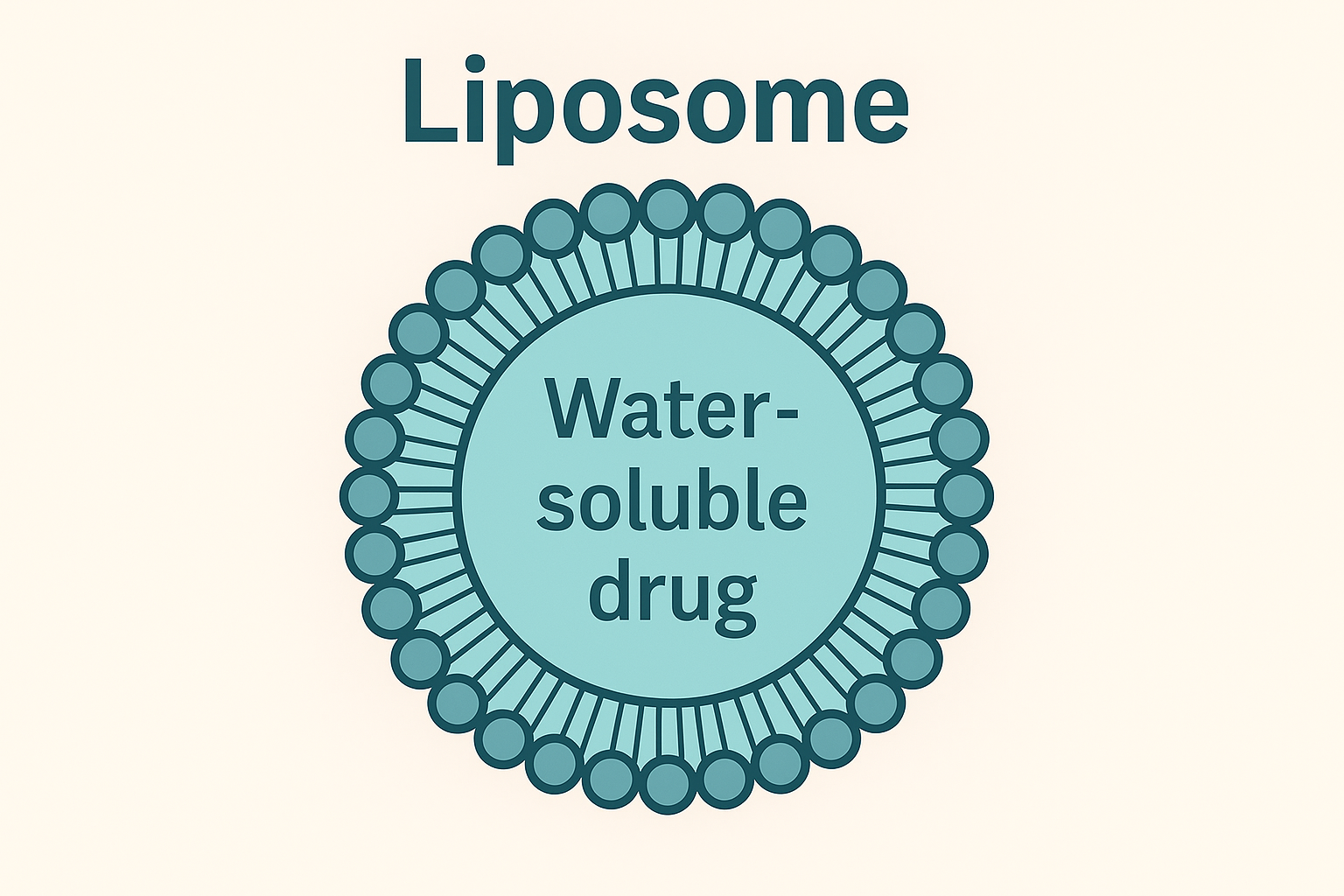
Ambisome® is a special formulation of amphotericin B. It uses tiny liposomes to deliver the drug.
What is a Liposome?
A liposome is a microscopic, fat-based bubble. It:
-
Protects the medicine until it reaches the right part of the body
-
Reduces irritation and side effects
-
Helps deliver amphotericin more gently to the lungs
You can think of liposomes like tiny protective vans, carrying the medicine where it’s needed most — often areas affected by CPA or ABPA.
Benefits of Inhaled Liposomal Amphotericin
-
Better tolerated than older versions (especially important for people with sensitive airways)
-
Safer for the lungs and kidneys
-
Can be used to target Aspergillus in the lungs directly
-
Suitable for people with CPA or difficult-to-control ABPA
-
May be used alongside antifungal tablets or corticosteroids
What to Expect During Treatment
-
You’ll use a nebuliser, a machine that turns liquid medicine into a fine mist.
-
Treatment usually takes around 15–30 minutes.
-
You may be asked to use a bronchodilator inhaler first (e.g. salbutamol) to open up your airways.
-
Your first treatment may be supervised to check for any side effects.
Common Side Effects
Most people tolerate liposomal amphotericin well, but possible side effects include:
-
Mild coughing or throat irritation
-
Chest tightness or wheezing (more likely with non-liposomal versions)
-
Unpleasant taste or dry mouth
People with ABPA may be more sensitive to these effects due to their underlying allergic response. If you have CPA, it’s important to report any new or worsening symptoms like increased coughing or breathlessness.
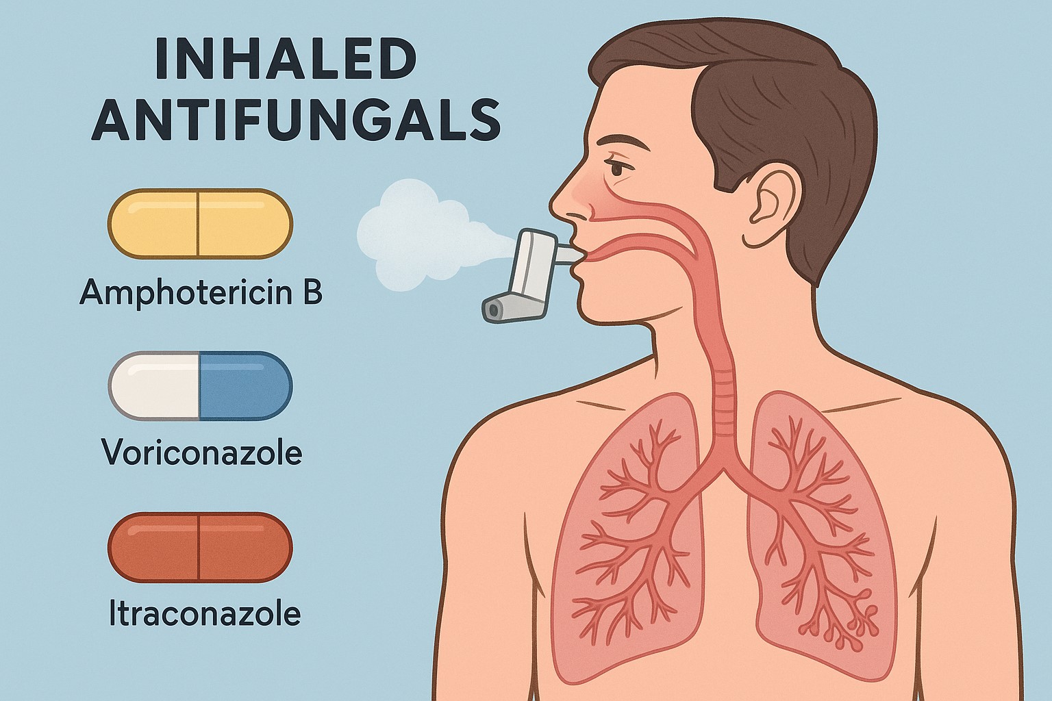
Inhalable antifungal medication for Aspergillosis
Inhaled antifungals are an area of active development, especially for targeting fungal lung infections like aspergillosis and candidiasis. This approach allows for high local drug concentrations in the lungs while minimizing systemic side effects. Here’s a summary of current and emerging inhaled antifungals:
✅ Currently Available or in Clinical Use (select cases or trials)
| Antifungal | Formulation | Indication / Use | Notes |
|---|---|---|---|
| Amphotericin B (liposomal) | Inhaled (off-label) | Prophylaxis in immunocompromised patients (e.g. post-transplant) | Used for inhaled prophylaxis against invasive aspergillosis; available in some UK centres |
| Voriconazole | Inhaled (compounded) | Limited use in chronic fungal lung disease | Very limited data; some use in compassionate settings |
| Itraconazole | Inhaled (experimental) | Chronic pulmonary aspergillosis | Inhalable versions have been studied (e.g. PUR1900/Pulmazole) |
| Nystatin | Inhaled (rare/off-label) | Oropharyngeal candidiasis or tracheobronchial use | Sometimes nebulized in ICU; limited absorption |
🧪 In Development / Clinical Trials
| Antifungal | Developer / Status | Target Use | Notes |
|---|---|---|---|
| Opelconazole (PC945) | Pulmocide Ltd – in Phase 3 trials | Inhaled for chronic aspergillosis, prophylaxis | Designed specifically for inhalation; long lung retention, minimal systemic exposure |
| Pulmazole (PUR1900) | Pulmatrix (partnering with Cipla) – early trials | ABPA, CPA in asthma/bronchiectasis | Inhaled itraconazole dry powder; promising lung targeting |
| Inhaled amphotericin B lipid complex | Aridis / others | Invasive fungal prophylaxis | Advanced animal and some early human data |
| Encochleated Amphotericin B | Matinas BioPharma (oral/inhaled being explored) | Aspergillosis, mucormycosis | Cochleate delivery protects drug; inhaled route under study |
🔬 Preclinical / Exploratory
| Antifungal Class | Notes |
|---|---|
| Echinocandins (e.g. caspofungin) | Not yet available in inhaled form, but being explored for nebulization |
| Azole reformulations | Research ongoing into nebulized posaconazole or isavuconazole for direct lung delivery |
| Novel agents (e.g. olorofim) | Olorofim is oral/IV only currently, but inhaled versions could emerge in future studies |
🧩 Potential Advantages of Inhaled Antifungals
-
High concentration directly at the site of infection (lungs)
-
Reduced systemic toxicity
-
Less interaction with hepatic CYP450 pathways (important for azoles)
-
Better for long-term suppression in CPA, ABPA, SAFS
🚧 Challenges
-
Delivery devices and patient technique (e.g. DPI vs nebuliser)
-
Ensuring adequate deposition in damaged or obstructed airways
-
Regulatory hurdles due to novel delivery routes
-
Limited real-world data so far
🌍 Fungal Spore Release in Different Climates
Climate has a major impact on fungal spore patterns. The types of fungi, timing of spore release, and health risks vary greatly between temperate, tropical, arid, and cold climates.
🌦️ Temperate Climates (e.g. UK, Northern Europe, Pacific Northwest USA)
-
Seasonal peaks: late summer to early autumn
-
Cladosporium, Alternaria, Aspergillus, Penicillium dominate
-
Triggers: humidity, rain, decaying vegetation, compost
-
Evening/night peaks for many spore types
🧠 Health risk: ABPA, asthma, SAFS, especially after garden work or composting
🌴 Tropical and Subtropical Climates (e.g. SE Asia, Central America, parts of Africa)
-
High year-round spore loads
-
Fungi: Aspergillus, Rhizopus, Mucor, Curvularia, Basidiomycetes, tropical molds
-
Daily rain cycles promote spore germination and release
-
Basidiospores and hyaline molds surge during evening and after rainfall
🧠 Higher incidence of fungal sinusitis, allergic fungal rhinosinusitis, and invasive infections in immunocompromised patients
🏜️ Arid and Semi-Arid Climates (e.g. Southwest USA, parts of the Middle East, Northern Africa)
-
Lower general spore burden, but dust storms can release huge amounts
-
Fungi: Coccidioides spp. (Valley fever), Aspergillus, dryland molds
-
Dry conditions favour small, dry spores that aerosolise easily
-
Wind and soil disturbance (construction, farming) are major triggers
🧠 Coccidioidomycosis, aspergillosis, dust-related asthma
❄️ Cold or Alpine Climates (e.g. Scandinavia, Alps, Canadian north)
-
Lower fungal biodiversity
-
Short seasonal spore peaks (spring–summer)
-
Indoor exposure more relevant: Penicillium, Cladosporium, Aspergillus
-
Mold growth occurs in poorly ventilated buildings (e.g. after snow melts)
🧠 Winter indoor mould exposure can worsen asthma, ABPA, SAFS
📌 Key Takeaway:
Different climates = different fungal threats.
The more humid, warm, or plant-rich the environment, the more active and diverse the fungal spore release — but indoor mould exposure is relevant in every climate.
Types of Window Screens for Allergen Protection
Yes — there are specialised window screens designed specifically to block allergens, including fungal spores, pollen, and dust. These can be a practical and effective solution for patients with ABPA, SAFS, asthma, CPA, or other respiratory allergies.
✅ 1. Pollen and Allergen Screens (Micromesh / Allergy Mesh)
-
Made from ultrafine polyester or polypropylene mesh
-
Pore size: typically 0.3–0.5 mm, small enough to block:
-
Pollen
-
Fungal spores (like Aspergillus and Cladosporium)
-
Insect particles
-
-
Allows air flow while reducing allergen load
🛒 UK Brands / Products to Look For:
-
Poll-Tex®: Proven in European studies to block >99% of airborne pollen and fine particles.
-
Neat Pleat® or Flyscreen UK Allergy Mesh
-
Tesa® Insect Stop Pollen Protection
-
Amazon or B&Q: Offer DIY-fit mesh kits
✅ 2. HEPA-Filtered Window Units
-
Some products combine an extractor fan + HEPA filter, ideal for:
-
Rooms needing airflow without outdoor contaminants
-
Urban or high-spore environments
-
-
More expensive but very effective
Examples:
-
Vent-Axia PureAir Room Ventilator
-
Aereco HRV or HEPA filter units
🛠️ Installation Tips
-
Choose removable or magnetic frames for rented homes
-
Ensure tight seal around edges to prevent gaps
-
Use with dehumidifiers or HEPA air purifiers indoors for extra control
💡 Bonus Tip:
-
Leave windows open early morning or midday if you must ventilate — avoid dusk/night when fungal spore levels rise.
-
Don’t open windows near compost heaps, sheds, or leafy overgrowth.
Managing Life with Haemoptysis
Managing life with haemoptysis — especially when it's recurrent, low-volume, or threatening to recur — can be physically and emotionally exhausting. Whether due to CPA, ABPA, bronchiectasis, aspergillus bronchitis, or other underlying lung conditions, the goal is to minimise triggers, support healing, and maintain safety without living in constant fear.
Here’s a comprehensive, practical guide to managing haemoptysis during recovery or periods of fragility:
🔴 Understanding the Risk
Haemoptysis (coughing up blood) can range from:
-
Minor (streaks in mucus)
-
Moderate (5–50ml)
-
Massive or life-threatening (>200–600ml in 24h — a medical emergency)
If you're in a recovery phase, you may be:
-
Post-bleed but still inflamed
-
Dealing with recurrent trickles
-
Worried about provoking a bleed due to fragile blood vessels or fungal activity
✅ Core Management Goals
| Goal | How to Achieve It |
|---|---|
| Prevent rebleeding | Avoid straining, irritating airways, or increasing pressure |
| Allow fragile vessels to heal | Stay well-hydrated, avoid airway trauma, reduce inflammation or infection |
| Identify and treat causes | Maintain antifungal, antibiotic or anti-inflammatory treatment as prescribed |
| Stay calm during symptoms | Know how to position yourself and who to contact |
| Keep life going gently | Pace activity, prioritise rest, manage anxiety without isolation |
🔹 1. Activity & Positioning: How to Move Safely
-
Avoid intense exercise, heavy lifting, straining (including on the toilet).
-
Keep your head elevated when sleeping (2 pillows or wedge).
-
If coughing blood:
-
Sit upright or lean slightly forward (don’t lie flat).
-
Lie on the side that’s bleeding (if known) — this protects the better lung.
-
🔹 2. Breath & Cough Management
-
Cough suppression may help reduce vessel trauma:
-
Use warm steam or gentle hydration first.
-
Use prescribed suppressants only if safe (some conditions need mucus clearance).
-
-
Huffing can be gentler than coughing.
-
Avoid dry air — use a humidifier, nasal rinses, or saltwater gargles.
🔹 3. Medication Adherence
-
Antifungals (e.g., voriconazole, itraconazole): Maintain strict levels.
-
Steroids (if prescribed): Taper cautiously under supervision.
-
Antibiotics or macrolides: Prevent secondary infection.
-
Tranexamic acid: Sometimes used short-term to reduce bleeding risk (under guidance).
🔹 4. Environmental & Lifestyle Support
-
Avoid dust, smoke, aerosols, strong odours, and temperature extremes.
-
Wear a mask when gardening, cleaning, or in crowded spaces.
-
Avoid alcohol and anti-inflammatory meds (NSAIDs) unless cleared.
-
Maintain gentle hydration, warm teas, and soothing soups to support healing.
🔹 5. Psychological Support: Managing Fear and Anxiety
-
It’s normal to fear rebleeding — but hypervigilance can increase stress and airway irritation.
-
Use mindfulness or grounding during panic (see above).
-
Talk with a support group or therapist if fear is affecting sleep or daily life.
-
Ask for a written plan from your medical team: “What to do if it happens again.”
🔹 6. When to Seek Help
Call your medical team or go to A&E if:
-
Bleeding increases or becomes bright red and continuous
-
You cough up >1 tablespoon of blood
-
You feel faint, breathless, or distressed
-
Blood is mixed with froth, or you feel it rising in your throat
📦 Preparedness Tips
-
Keep a rescue plan printed or saved on your phone.
-
Store clean cloths or tissues, bottled water, and calming items near where you rest.
-
Carry a medical ID or information card if you’re going out alone.
🧘♀️ Living Well While Letting It Heal
-
Maintain light, slow routines – a little movement, some fresh air, safe distraction.
-
Avoid cough triggers like strong smells or cold air.
-
Use the time to focus on recovery, build strength gradually, and connect with others.
🧠 What Is Mindfulness? Can it Help Living With Aspergillosis?
Mindfulness can be a powerful support tool for people living with aspergillosis, whether it's CPA, ABPA, SAFS, or related conditions like bronchiectasis or asthma. It doesn’t replace medical treatment, but it can significantly help with coping, symptom awareness, and emotional wellbeing.
Below is a breakdown of how mindfulness practices may benefit people with aspergillosis:
Mindfulness means paying attention, on purpose, in the present moment, without judgment. It involves noticing your thoughts, feelings, and physical sensations — without getting caught up in them or trying to change them immediately.
This can be done through:
-
Breath awareness
-
Body scans
-
Guided meditation
-
Mindful movement (e.g. yoga, tai chi)
-
Mindful walking or eating
🌿 How Can Mindfulness Help with Aspergillosis?
| Challenge | How Mindfulness Helps |
|---|---|
| Breathlessness / coughing fits | Helps calm panic and reduce over-breathing; improves awareness of breath patterns |
| Medication side effects (e.g. voriconazole) | Reduces anxiety and fear responses; helps observe sensations without spiralling |
| Chronic fatigue / brain fog | Supports pacing and acceptance; improves focus and attention |
| Pain or chest discomfort | Helps reduce distress by observing pain without fighting it (used in pain management) |
| Anxiety or health fear | Teaches how to sit with fear without letting it dominate thinking |
| Frustration with slow progress | Encourages self-kindness and reduces reactivity to setbacks |
| Isolation / low mood | Builds resilience and reduces rumination by shifting focus to what is happening now |
| Poor sleep | Calms a racing mind before bedtime |
🧘♀️ Simple Mindfulness Practices for Patients
1. Breath Awareness (1–2 minutes)
Focus gently on the in-breath and out-breath. When your mind wanders (and it will), gently bring it back to the breath.
Use when: You feel overwhelmed, panicked, or need to slow down.
2. Body Scan (5–10 minutes)
Lie or sit down, and slowly move your attention through your body from feet to head. Notice sensations — tension, warmth, numbness — without judgment.
Use when: Resting, falling asleep, or managing pain or fatigue.
3. Mindfulness in Everyday Tasks
Choose a task (e.g., washing up, brushing teeth, folding clothes). Do it slowly and notice the sensations, smells, sounds — staying fully present.
Use when: You feel disconnected or anxious during the day.
4. RAIN Method for Anxiety
-
Recognise what’s happening
-
Allow the experience to be there
-
Investigate with curiosity (Where in my body do I feel this?)
-
Nurture (What do I need right now?)
Use when: You’re feeling a strong emotional response (fear, frustration, sadness).
💬 What Patients Have Said
“It doesn’t stop the coughing, but I panic less when I feel it coming on.”
“I used to fear every twinge in my chest. Mindfulness gave me space before I reacted.”
“When I feel the visual side effects starting, I breathe and tell myself: just observe, it will pass.”
📱 Resources and Apps (Many Are Free)
-
Headspace (good for beginners)
-
Insight Timer (lots of free guided meditations)
-
Calm (for sleep and anxiety)
-
Breathworks (mindfulness for health conditions)
-
Smiling Mind (free and evidence-based)
⚠️ Important Notes
-
Mindfulness is a skill — it takes time and practice. Start small.
-
It can feel uncomfortable at first, especially if you're used to avoiding difficult thoughts or sensations.
-
If you’ve experienced trauma or distressing symptoms, guided mindfulness with a therapist may be safer.
Grounding Techniques for Aspergillosis: Coping with Panic, Anxiety, and Sensory Overload
Living with aspergillosis — whether it’s ABPA, CPA, SAFS, or another form — can be physically and emotionally overwhelming. From managing symptoms like breathlessness or cough, to coping with powerful medications (like voriconazole), or dealing with uncertainty and isolation, it’s not unusual to feel anxious, panicked, or disconnected.
Grounding techniques are simple, effective strategies that help you regain a sense of calm, control, and presence, even in the middle of a crisis.
🧭 What Are Grounding Techniques?
Grounding techniques are ways to anchor yourself in the present moment, especially when your mind or body feels out of control. They use your senses, movement, and breath to shift focus away from distressing thoughts, frightening physical sensations, or overwhelming emotion.
They are widely used by people living with:
-
Chronic illness
-
Anxiety or panic
-
Medication side effects (e.g. visual disturbances from voriconazole)
-
Post-traumatic stress
-
Neurological symptoms or dissociation
🩺 When Might a Person with Aspergillosis Use Grounding?
| Situation | How Grounding Can Help |
|---|---|
| Visual disturbances from medications like voriconazole | Reduce fear, reconnect with reality |
| Panic or anxiety about symptoms or breathlessness | Calm racing thoughts and slow the body’s stress response |
| Overstimulation in hospitals or crowded places | Help regulate sensory overload |
| Before or after medical appointments | Reduce anticipatory anxiety or recover afterward |
| During flare-ups, pain, or coughing episodes | Stay focused, steady, and more in control |
| While trying to sleep | Settle a restless mind or racing heart |
| When feeling isolated, overwhelmed, or low | Feel more present and safe in the body and environment |
✅ Simple Grounding Techniques to Try
🔹 1. The 5-4-3-2-1 Technique (Sensory Anchoring)
Name:
5 things you can see
4 things you can touch
3 things you can hear
2 things you can smell
1 thing you can taste
When to use: During a visual disturbance, panic, or if you feel disconnected.
🔹 2. Box Breathing
Inhale – 4 seconds
Hold – 4 seconds
Exhale – 4 seconds
Hold – 4 seconds
When to use: During a flare-up, medication side effect, or while waiting in a stressful situation (e.g., hospital, travel, scans).
🔹 3. Cold Sensation Reset
-
Hold an ice cube
-
Splash cold water on your face
-
Use a cooling gel pack on your hands or wrists
When to use: To snap out of panic or rapid heart rate, especially if you feel faint or detached.
🔹 4. Movement and Pressure
-
Press your feet firmly into the floor
-
Grip the arm of a chair or ball your fists and release
-
Walk slowly, focusing on each step
When to use: When anxious, overwhelmed, or before a test, clinic visit, or treatment.
🔹 5. Safe Words & Orientation
Say aloud:
“I’m [your name].
I’m in [location].
It’s [day/time].
I feel this way because [e.g. the medication is affecting me], but it will pass.”
When to use: After waking up anxious, during episodes of confusion, visual disturbance, or feeling “spaced out.”
🔹 6. Grounding Object or Support Contact
-
Carry a smooth stone, photo, textured card, or essential oil roller.
-
Call or message someone you trust.
-
Look at a comforting image or mantra you keep nearby.
When to use: When you need emotional reassurance or support — especially after a scary experience.
💡 A Note on Voriconazole Visual Effects
Visual side effects from voriconazole (e.g. flashing lights, color changes, or visual trails) usually:
-
Start within 30 min to 2 hours of a dose
-
Last 2 to 6 hours
-
Fade over time or after stopping the drug
Grounding techniques can’t stop the visual changes but can help you stay calm and reduce the fear or panic response they often trigger.
If the symptoms become unmanageable, let your clinical team know — they may adjust your dose or change the medication.
💬 Final Thoughts
Grounding techniques are safe, non-invasive, and free. They don’t cure symptoms but can help you cope more confidently, especially when dealing with the emotional and sensory impact of life with aspergillosis.
Many patients find it helpful to practice them regularly, even when they feel fine — so they’re easier to use in a crisis.
Visual Disturbances on Voriconazole: What’s Happening and Why It Can Feel So Scary
If you're experiencing frightening or panicked feelings due to visual disturbances while taking voriconazole, you're not alone — and there are reasons behind both the symptoms and your emotional response.
🧠 What Causes Visual Disturbances with Voriconazole?
-
Voriconazole crosses the blood–brain barrier and affects the central nervous system (CNS).
-
It interacts with retinal photoreceptors (especially rods), which may explain:
-
Lights seeming too bright or flickering.
-
Halos, flashes, or color distortions.
-
Visual “trails” following movement.
-
-
Although not fully understood, it may involve:
-
Temporary interference with visual signal processing in the brain and retina.
-
Altered neurotransmitter activity or enhanced retinal light sensitivity.
-
⏱️ When Do These Disturbances Happen and How Long Do They Last?
-
Onset: Symptoms typically begin 30 minutes to 2 hours after a dose.
-
Duration: They usually last 2 to 6 hours, fading as the drug is processed.
-
Resolution: Most people find the effects wear off before the next dose.
-
Over time, even if you stay on the drug, your body often adapts, and the symptoms lessen or disappear entirely within a few days to weeks.
😰 Why Do They Feel So Scary?
-
The effects are sudden and intense, often catching people off guard.
-
Our brains rely on visual input for safety — when this gets disrupted, it can trigger anxiety or panic.
-
If you live with anxiety or take other medications, your brain may amplify the sense of threat, even though the effect is temporary.
-
Descriptions like “psychedelic” or “derealised” are common — which understandably causes distress.
🩺 What Can You Do?
-
Talk to your medical team. They may:
-
Check voriconazole blood levels.
-
Reduce the dose or change timing.
-
Switch to an alternative antifungal if needed.
-
-
Avoid driving at night or doing visually demanding tasks until things settle.
-
If anxiety is a problem:
-
Try grounding techniques (e.g. breath control, sound orientation).
-
Ask your doctor whether temporary support (like anti-anxiety medication or CBT) might help.
-
-
Some patients find taking voriconazole in the evening lets them sleep through the worst of it — but check this with your prescriber first.
🔔 When to Get Urgent Help
If symptoms last unusually long, worsen over time, or include confusion, agitation, or hallucinations, contact your healthcare provider. These may be signs of CNS toxicity, especially if blood levels are high.
🧠 Why Some Medications Can't Be Prescribed by GPs

In the UK, the NHS uses a tiered prescribing system that sometimes prevents GPs from prescribing certain medications, even if those medicines are available elsewhere in the NHS.
Here’s a clear explanation of how and why this happens:
🔒 1. Shared Care or Specialist-Only Medications
Some medicines are designated as “specialist-only” or “shared care” treatments. This means:
-
GPs are not authorised to initiate them.
-
In some cases, they can continue a prescription once a specialist starts it — but only if a formal shared care agreement is in place.
Examples include:
-
Biologics for asthma, ABPA, or autoimmune disease
-
High-risk antifungals like voriconazole or posaconazole
-
Certain cancer, transplant, or hormone drugs
This system ensures that:
-
The medication is closely monitored by someone with specialist knowledge
-
Risks like interactions, side effects, and required blood tests are safely managed
📜 2. Local Prescribing Formularies
Each NHS Integrated Care Board (ICB) or local NHS Trust maintains a formulary — a list of medicines approved for use in that area.
-
If a medicine isn't on the local formulary, the GP may be unable to prescribe it, even if NICE (the National Institute for Health and Care Excellence) says it's effective.
-
These decisions are based on local budget priorities, agreements with hospitals, and clinical capacity.
💷 3. Cost Controls and Prior Approvals
Some medications are expensive or highly specialised, and require:
-
Prior approval by a funding panel
-
A hospital-based consultant to apply for and justify the treatment
GPs usually cannot access these approval pathways directly.
⚠️ 4. Liability and Risk
Even if a GP understands the condition, they may not have:
-
Access to monitoring protocols
-
Up-to-date knowledge of rare drug interactions or side effects
-
The ability to interpret complex blood results needed for safe prescribing
For legal and safety reasons, GPs must follow guidance from their local ICB or NHS England on what they can and can’t prescribe.
✅ What Patients Can Do
-
Ask the hospital team if the medication can be prescribed under shared care, and whether your GP has agreed to it.
-
Ask your GP to request guidance from the local medicines management team.
-
Request a hospital prescription if urgent — but note this often requires collection from hospital pharmacies.