
Can blood tests help predict if chronic pulmonary aspergillosis will come back?
This study from the National Aspergillosis Centre (NAC) looked at people with chronic pulmonary aspergillosis (CPA) who had completed antifungal treatment and asked a simple question:
Can blood tests tell us who is more likely to relapse after treatment stops?
What the researchers did
Doctors reviewed patients with CPA who had:
-
Taken antifungal treatment for at least 6 months
-
Stopped treatment because they were clinically stable
They then followed these patients to see who stayed well and who relapsed, and compared this with their blood test results at the time treatment stopped.
What they found
-
About 1 in 4 patients had a relapse after stopping treatment
-
People whose Aspergillus IgG blood test was still high at the end of treatment were much more likely to relapse
-
Patients whose IgG level had fallen to a lower level did not relapse in this study
-
Signs of Aspergillus allergy or sensitisation also increased relapse risk
-
CT scan appearances and treatment length alone were not reliable predictors
Why this matters for patients
This means that:
-
Blood tests may help doctors decide when it is safe to stop treatment
-
Some people may need closer follow-up or longer treatment
-
Follow-up can be more personalised, rather than “one size fits all”
Importantly, a relapse does not mean treatment failed — it reflects how persistent this infection can be in damaged lungs.
Key takeaway
A simple blood test at the end of treatment may help predict who needs closer monitoring for CPA relapse.
This research supports a more individualised approach to long-term CPA care.
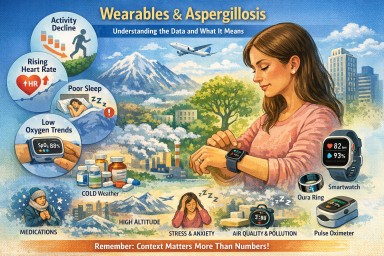
Wearable devices and aspergillosis
Are they useful yet – and which ones are the most accurate?
The short answer
Wearable devices do not diagnose aspergillosis and cannot tell what is causing symptoms.
However, some wearables are now good enough to provide useful background information about how your body is coping over time.
Their value lies in:
-
spotting gradual deterioration
-
recognising patterns over weeks or months
-
supporting conversations with your clinical team
They are not a replacement for scans, blood tests, sputum cultures, lung function tests, or specialist review.
What wearables can realistically help with
For people with:
-
Chronic Pulmonary Aspergillosis (CPA)
-
Allergic Bronchopulmonary Aspergillosis (ABPA)
-
Aspergillus bronchitis
-
Aspergillosis with bronchiectasis or asthma
wearables can sometimes help answer:
-
“Am I slowly getting worse, or is this just a bad patch?”
-
“Has my recovery from exertion changed?”
-
“Are my nights becoming more disrupted?”
They are most useful for long-term trends, not day-to-day decisions.
The signals that matter most
From both patient experience and respiratory clinical practice, these signals tend to be most meaningful:
1. Activity tolerance
-
Falling step count over weeks
-
Needing longer to recover after usual activity
-
Avoiding activity you previously managed
➡ Often one of the earliest signs of deterioration.
2. Resting heart rate
-
A persistent rise from your own baseline
-
Especially if not explained by infection, fever, medication or stress
➡ Often reflects physiological strain before symptoms become obvious.
3. Sleep quality
-
Frequent night waking
-
Shortened or fragmented sleep
-
Feeling unrefreshed despite enough hours in bed
➡ Poor sleep often accompanies worsening respiratory symptoms or medication effects.
4. Oxygen saturation (SpO₂) trends
-
Repeated low readings
-
Drops overnight or with exertion
-
Patterns that persist over days or weeks
➡ Trends matter far more than single readings.
➡ Dedicated oxygen monitors are usually more reliable than watches.
What about ECG and breathing rate?
These features are often misunderstood. They are not useless, but they are supportive rather than central in aspergillosis care.
ECG (heart rhythm)
Some wearables can record a single-lead ECG, which may detect:
-
atrial fibrillation
-
sustained rhythm abnormalities
This can be helpful if someone develops:
-
new palpitations
-
breathlessness out of proportion to lung symptoms
-
dizziness or faintness
➡ ECG does not provide information about Aspergillus activity or lung disease progression.
Breathing (respiratory) rate
Most wearables estimate breathing rate indirectly, usually during sleep.
Breathing-rate trends may:
-
support a sense that breathing effort has increased
-
highlight disrupted sleep linked to respiratory load
➡ It cannot distinguish fungal disease from asthma, infection, anxiety or medication effects.
Medication and age matter — a lot
When clinicians interpret wearable data, they always consider:
-
antifungal medicines (e.g. azoles)
-
steroids (current or past)
-
asthma and allergy treatments
-
other long-term conditions
-
age-related physiological change
Common medication effects seen on wearables
-
higher resting heart rate
-
poorer sleep
-
fatigue
-
reduced activity tolerance
These are common and expected and do not automatically mean disease progression.
As we age:
-
recovery slows
-
sleep becomes lighter
-
heart-rate variability reduces
-
oxygen dips more easily overnight
➡ Always compare data to your own baseline, not to “normal” values.
Environment and everyday factors strongly affect readings
Often more than lung disease itself
This makes them highly sensitive to environment and daily circumstances.
Many “abnormal” readings reflect conditions around you, not worsening aspergillosis.
Temperature (especially cold)
Cold causes blood vessels in the skin to narrow, which can lead to:
-
falsely low oxygen readings
-
erratic heart-rate data
-
missing or failed measurements
Common situations:
-
cold bedrooms
-
winter walks
-
sleeping with arms outside the duvet
➡ A low oxygen reading in the cold is often technical, not medical.
Altitude and air pressure
At higher altitude (even modest):
-
oxygen saturation normally falls
-
breathing rate may rise
-
sleep may worsen
Examples:
-
flying
-
holidays in hilly or mountainous areas
-
high-rise accommodation
➡ This is normal physiology, not disease progression.
Air quality, humidity and heat
Poor air quality or high humidity can cause:
-
faster breathing
-
increased heart rate
-
worse sleep
-
reduced activity tolerance
➡ Wearables detect body stress, not its cause.
Sleep environment
Sleep data is very sensitive to:
-
noise
-
light
-
room temperature
-
uncomfortable bedding
A poor sleep score often reflects environmental disruption, not lung decline.
Movement, posture and coughing
Night-time data can be affected by:
-
coughing
-
restless sleep
-
sleeping on the arm wearing the device
➡ Night data is often noisy and imperfect.
Hydration, alcohol and meals
-
dehydration → higher heart rate
-
alcohol → worse sleep and altered breathing rate
-
heavy evening meals → raised heart rate
These effects are temporary and not signs of deterioration.
Stress and anxiety
Stress can:
-
raise heart rate
-
increase breathing rate
-
worsen sleep
Wearables cannot distinguish stress from illness, and worrying about readings can make readings worse — a common feedback loop.
What wearables cannot do (important)
-
They cannot diagnose aspergillosis
-
They cannot identify fungal flares
-
They cannot separate cause from effect
-
They cannot replace specialist investigations
They provide context, not answers.
The most accurate consumer devices (2025–26)
Best overall smartwatches
-
Apple Watch (Series 9 / Ultra 2) – excellent heart-rate accuracy, ECG, good sleep trends
-
Withings ScanWatch 2 – health-focused, ECG and oxygen, long battery life
-
Garmin Venu 3 / Epix Pro – excellent activity and recovery tracking
Best non-watch wearables
-
Oura Ring (Gen 3) – strong overnight physiology and sleep trends
-
Wellue O2Ring / similar continuous oximeters – more reliable oxygen trends than watches
These are listed because of better accuracy and consistency, not because they are diagnostic devices.
Can wearable data cause over-worry?
Yes — and this is common, especially in people with long-term lung disease.
Wearables can sometimes:
-
increase anxiety
-
encourage constant checking
-
turn normal variation into worry
-
make people feel unwell even when stable
This is not a personal weakness.
When wearables help
-
checked occasionally
-
viewed over weeks or months
-
used to support (not replace) symptoms
When wearables stop helping
-
if they increase anxiety
-
if they disrupt sleep
-
if numbers override how you feel
➡ It is entirely reasonable to reduce use or stop.
How specialists actually prioritise information
In real aspergillosis care, clinicians still focus on:
-
How you feel
-
What you can do
-
Symptoms and sputum
-
Imaging and tests
-
Medication history
Wearable data sits well below these.
The bottom line
-
✔ Wearables are becoming useful for monitoring trends
-
✔ ECG and breathing rate add context and safety, not answers
-
✔ Medication, age and environment strongly affect readings
-
❌ Wearables do not diagnose aspergillosis
-
✔ If a device increases anxiety, stepping back is sensible
Antifungal Medicines: Dosing, Monitoring, and the Role of Specialist Care
A detailed reference for patients and non-specialist clinicians
1. Why antifungal treatment is different from most medicines
Oral antifungal medicines—especially azole antifungals—are essential for treating long-term fungal diseases such as chronic pulmonary aspergillosis and allergic bronchopulmonary aspergillosis.
They differ from many common medicines because they:
-
Have a narrow margin between effectiveness and toxicity
-
Behave very differently between individuals
-
Are often taken for months or years, not days
-
Interact with many commonly prescribed drugs
For these reasons, antifungal treatment requires individualised dosing, monitoring, and specialist input, rather than a standard fixed dose.
2. What “pharmacokinetics” means (plain language)
Pharmacokinetics describes what the body does to a drug:
-
Absorption – how well the drug enters the bloodstream from the gut
-
Distribution – how effectively it reaches tissues such as the lungs
-
Metabolism – how quickly the liver breaks it down
-
Elimination – how the drug leaves the body
Differences at any of these stages explain why the same dose can be ineffective for one person and toxic for another.
3. Different generations of azole antifungals behave differently
Each generation of azole antifungal was designed to improve effectiveness, but chemical changes also altered how the body handles the drug.
First-generation azoles (older drugs)
Examples
-
Ketoconazole
-
Fluconazole (limited activity against Aspergillus)
Key features
-
Variable absorption
-
Shorter half-life
-
Less reliable lung penetration
Clinical relevance
-
Rarely used now for chronic aspergillosis
Second-generation azoles (mainstay treatment)
Examples
-
Itraconazole
-
Voriconazole
-
Posaconazole
Key features
-
Excellent lung and tissue penetration
-
Highly variable metabolism between people
-
Strong interaction with liver enzymes
Clinical relevance
-
Very effective
-
Blood levels vary widely
-
Dose adjustment and monitoring are often essential
Newer azoles
Example
-
Isavuconazole
Key features
-
More predictable absorption
-
Long, stable half-life
-
Fewer extreme peaks and troughs
Clinical relevance
-
Often better tolerated long-term
-
Monitoring still important, but dosing may be more stable
4. Why the “right dose” matters so much
Too little antifungal
-
Infection not adequately controlled
-
Symptoms persist or worsen
-
Risk of antifungal resistance
-
Fewer future treatment options
Too much antifungal
-
Liver irritation or damage
-
Nausea, appetite loss
-
Neurological or visual side effects
-
Drug accumulation, especially with long-term use
The aim is always the lowest dose that effectively controls the fungus.

5. How clinicians know whether the dose is right
No single test determines this. The correct dose is identified when three elements align:
1️⃣ Blood level testing (therapeutic drug monitoring)
-
Measures how much drug is actually in the bloodstream
-
Helps identify:
-
Under-dosing
-
Target-range dosing
-
Toxic levels
-
2️⃣ Clinical response
-
Symptoms stabilise or improve
-
Fewer flare-ups or complications
-
Better day-to-day function
3️⃣ Safety monitoring
-
Liver and kidney blood tests
-
Review of side effects
-
Ongoing assessment of drug interactions
Only when effectiveness and safety are both acceptable is the dose considered “right”.
6. Why the right dose can change over time
A dose that was correct initially may later need adjustment because of:
-
Weight or body-composition changes
-
Age-related metabolic changes
-
New medications (including antibiotics or steroids)
-
Changes in liver or kidney function
-
Gradual drug accumulation during long-term therapy
Regular review is therefore expected and appropriate.
7. Is it sometimes impossible to find a stable dose?
Yes. For a minority of patients, a perfectly balanced dose cannot be found.
Reasons include:
-
Extremely fast or slow drug metabolism
-
A very narrow safety window
-
Long-term toxicity despite “acceptable” blood levels
-
Unavoidable interacting medications
-
Liver, kidney, or neurological vulnerability
-
Partial or full antifungal resistance
In these cases, the dose that controls the fungus and the dose that causes side effects may overlap.
This reflects biological limits, not treatment failure.
8. What clinicians do when a stable dose cannot be achieved
Options may include:
-
Switching to a different azole with different pharmacokinetics
-
Using modified dosing schedules (split dosing, slower titration)
-
Accepting a lower suppressive dose rather than full eradication
-
Considering non-azole antifungals where appropriate
-
Prioritising symptom control and quality of life
All are intentional, safety-focused decisions.
9. The central role of the specialist pharmacist
Specialist pharmacists are key to safe antifungal care, particularly for long-term azole therapy.
They play a critical role in:
Interpreting drug levels
-
Assessing whether a level is truly low or high
-
Accounting for dose timing and formulation
-
Preventing unnecessary or unsafe dose changes
Managing drug–drug interactions
Azoles interact with many common medicines, including:
-
Steroids and inhalers
-
Heart rhythm drugs
-
Blood thinners
-
Anti-epileptics
-
Pain medications
The specialist pharmacist:
-
Reviews the full medication list
-
Anticipates interactions before harm occurs
-
Advises on adjusting both interacting drugs
Individualising dosing
When standard doses do not work, they help design:
-
Non-standard doses
-
Split dosing schedules
-
Slow titration plans
-
Alternative azoles with different pharmacokinetics
Protecting patients during long-term treatment
They monitor:
-
Trends in liver and kidney tests
-
Signs of cumulative toxicity
-
Whether symptoms may be drug-related rather than disease-related
Coordinating care
They act as a bridge between:
-
Laboratory results
-
Clinical decision-making
-
Patient experience

Their involvement often changes management, not just fine-tunes it.
10. Where antifungal drug level testing is done in the UK
In the UK, antifungal drug level testing is centralised.
-
Blood samples are taken locally
-
Samples are sent to specialist reference laboratories, most commonly the
Mycology Reference Centre Manchester -
Results are returned to the local clinical team for interpretation
Patients managed through specialist services such as the
National Aspergillosis Centre
benefit from integrated expertise in antifungal pharmacology, imaging, and long-term monitoring.
This process is routine and standard for antifungal care.
11. Key reassurance for patients
-
Dose changes are normal and expected
-
Side effects are often biology-driven, not your fault
-
Blood tests make treatment safer, not riskier
-
Switching drugs is a planned strategy, not giving up
12. One-paragraph summary
Antifungal medicines—particularly azole antifungals—have complex and highly variable behaviour in the body, with a narrow balance between effectiveness and toxicity. Safe use requires individualised dosing, therapeutic drug monitoring, symptom review, and long-term safety checks. Specialist pharmacists play a central role in interpreting drug levels, managing interactions, and tailoring treatment. For some patients, a perfectly balanced dose cannot be achieved, and alternative strategies are required. This reflects biological complexity, not failure, and the overarching aim is always effective fungal control with the best possible long-term safety and quality of life.

New UK Best Practice Guidelines for Diagnosing Fungal Diseases – What Patients Need to Know
A major new set of UK guidelines has just been published on how doctors and laboratories should diagnose serious fungal diseases. These come from the British Society for Medical Mycology (BSMM) and aim to make diagnosis faster, more accurate, and more consistent across the country.
For patients and families, this is very good news.
Why does this matter?
Fungal diseases can be difficult to diagnose. Many symptoms overlap with other conditions, and traditional tests sometimes miss infections. This can lead to delays, uncertainty, or unnecessary treatments.
The new BSMM recommendations help make sure that:
-
The right tests are used at the right time
-
Results come back more quickly
-
Hospitals know which modern tests give the best answers
-
Doctors can decide sooner whether antifungal treatment is needed
-
Unnecessary or ineffective treatments can be avoided
Overall, this means quicker diagnoses, fewer missed cases, and better care.
What is new in these guidelines?
The guidance highlights several improvements that directly benefit patients:
1. More accurate tests
Doctors are encouraged to use modern tests such as PCR, antigen tests, and antibody tests—these can detect fungal infections earlier and more reliably than traditional culture alone.
2. Faster turnaround times
Hospitals are encouraged to report important results within hours, not days. Faster answers mean faster treatment.
3. Better testing for people with chronic lung conditions
People with asthma, bronchiectasis, COPD, cystic fibrosis, or other long-term lung problems are now recognised as groups who may need access to fungal testing sooner.
4. Clearer pathways for difficult-to-diagnose conditions
Conditions such as chronic pulmonary aspergillosis (CPA), allergic bronchopulmonary aspergillosis (ABPA), fungal bronchitis and invasive fungal infections now have clearer testing strategies.
5. Stronger links to antifungal stewardship
This means using antifungal medicines only when they are truly needed, helping prevent side-effects and resistance.
6. Guidance for hospitals with fewer resources
The document includes a step-by-step approach to help smaller or overseas hospitals improve gradually.
What does this mean for patients?
If you are living with, or being assessed for, a fungal condition, you should expect:
-
More consistent tests wherever you are treated in the UK
-
Better access to specialist testing when needed
-
Earlier and more confident diagnoses
-
More appropriate treatments, with less trial-and-error
-
Closer monitoring during treatment
These improvements could reduce anxiety, cut down on repeated appointments, and help your clinical team make clearer decisions.
Who should be following these guidelines?
-
Hospital laboratories
-
Doctors and nurses in respiratory, ICU, oncology, infectious diseases, and transplant services
-
GP practices referring patients for investigation
-
Healthcare commissioners
-
Hospitals outside the UK wanting to improve fungal care
Patients and carers can also play a part by asking:
“Does my hospital follow the latest BSMM recommendations for fungal testing?”
The bottom line
These new BSMM guidelines are a major step forward for anyone affected by fungal disease. They promote earlier diagnosis, better access to testing, safer treatment, and improved outcomes.
Putting these recommendations into everyday practice—across the UK and worldwide—has the potential to transform care and save lives.
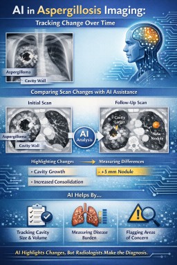
The role of AI in imaging follow-up for aspergillosis
Aspergillosis is not a single disease, but a spectrum of related conditions, including allergic bronchopulmonary aspergillosis (ABPA), chronic pulmonary aspergillosis (CPA), aspergilloma, subacute invasive aspergillosis, and Aspergillus bronchitis. Across this spectrum, imaging plays a central role in assessment, monitoring, and treatment planning.
Artificial intelligence (AI) is increasingly being explored in chest imaging as a supportive tool, particularly in the follow-up of chronic and complex lung disease. Its potential value in aspergillosis lies not in making diagnoses, but in consistent, data-driven image comparison.
Why aspergillosis is challenging for imaging interpretation
Imaging findings in aspergillosis are often:
-
Non-specific, overlapping with tuberculosis, non-tuberculous mycobacterial lung disease, lung cancer, vasculitis, and post-infectious scarring
-
Heterogeneous, even within the same patient
-
Slowly evolving, making change difficult to judge by eye
-
Mixed with other lung disease, such as bronchiectasis, asthma, fibrosis, or emphysema
For these reasons, imaging must always be interpreted in full clinical context and often benefits from specialist multidisciplinary discussion.
Where AI may add particular value
AI does not “know” that it is looking at aspergillosis. Instead, it compares imaging data directly, without assumptions about diagnosis. This can be helpful in several areas relevant to aspergillosis:
1. Consistent comparison of serial scans
AI can compare computed tomography (CT) scans over time using the same criteria each time, helping to:
-
Detect subtle interval change in cavities or nodules
-
Measure changes in cavity size, wall thickness, or internal content
-
Identify progression or stability that may be difficult to judge visually
This is particularly useful in chronic pulmonary aspergillosis, where progression may be slow and subtle.
2. Objective measurement of disease burden
AI can assist with:
-
Quantifying cavity volume or consolidation
-
Measuring extent of bronchiectasis or mucus plugging
-
Tracking changes following antifungal treatment or airway clearance
Objective measurements may help reduce subjectivity when monitoring response to treatment.
3. Highlighting areas for closer review
AI systems can flag areas of change or abnormality for radiologist attention. This may act as a “second set of eyes”, particularly in busy services, but does not replace expert review.
What AI cannot do in aspergillosis
It is important to be clear about the limitations:
-
AI cannot diagnose aspergillosis
-
AI cannot distinguish colonisation from active disease
-
AI cannot integrate symptoms, immune status, serology, or microbiology
-
AI cannot judge clinical significance or treatment need
For example, a change in cavity appearance may reflect active disease, treatment response, bacterial infection, bleeding, or simple movement of intracavitary material. Only expert clinical interpretation can determine significance.
Why radiologist expertise remains essential
In aspergillosis, small imaging changes can have very different meanings depending on context. Radiologists bring:
-
Experience in recognising mimics and artefacts
-
Understanding of treatment-related change
-
Ability to communicate uncertainty and recommend next steps
-
Integration of imaging with wider clinical information
AI may improve consistency and sensitivity, but responsibility for interpretation and reporting remains with the radiologist.
A balanced way to think about AI in aspergillosis imaging
In aspergillosis, artificial intelligence is best viewed as a tool that highlights and measures change, rather than one that explains or diagnoses it. Its strength lies in consistency; its limitation lies in lack of clinical understanding.
Used appropriately, AI may support safer and more consistent follow-up for people living with aspergillosis, while expert radiology and specialist clinical care remain central to diagnosis and management.

Using Radiopaedia and Online Imaging Resources Safely: What Expert Patients and Non-Specialist Clinicians Need to Know
Online radiology education platforms such as Radiopaedia (see aspergillosis images here) have transformed access to medical knowledge. They provide high-quality explanations of imaging findings, annotated examples, and differential diagnoses that are invaluable for learning, teaching, and patient empowerment.
For expert patients living with long-term conditions, and for non-specialist clinicians working outside radiology, these resources can greatly improve understanding of scan reports and discussions with healthcare teams. However, it is important to understand what these tools can – and cannot – do.
Radiopaedia is an educational resource, not a diagnostic service
Radiopaedia is designed to teach pattern recognition and radiological reasoning, not to provide individual diagnoses. The cases shown are curated examples, often with classic features, and are presented without the full clinical complexity that accompanies real patients.
Real-world imaging interpretation requires integration of:
-
Clinical history and symptoms
-
Laboratory results (for example inflammatory markers, microbiology, immunology)
-
Prior imaging and disease progression
-
Treatment history and response
-
Knowledge of common mimics and incidental findings
This clinical synthesis cannot be replicated by reviewing example images alone.
Why expert radiologist review still matters
For many diagnoses, there is no substitute for a radiologist formally reviewing and interpreting the imaging.
This is particularly true when:
-
Findings are subtle or evolving
-
Multiple conditions coexist (for example bronchiectasis, infection, scarring, and inflammation together)
-
Imaging appearances overlap between diseases
-
Treatment decisions depend on small but important changes over time
Radiologists are trained to recognise not only “textbook” appearances, but also atypical, incomplete, or misleading patterns, and to weigh uncertainty appropriately in their reports.
Imaging patterns are rarely diagnostic in isolation
Many imaging features are non-specific. For example:
-
Cavities can be caused by infection, inflammation, malignancy, or prior disease
-
Nodules may represent infection, scarring, inflammation, or benign change
-
Mucus plugging can occur in asthma, infection, allergic disease, or chronic airway disease
Educational resources often present differential diagnoses clearly, but deciding which diagnosis applies to a specific patient requires clinical judgment and experience.
A particular note for chronic lung and fungal disease
In complex conditions such as chronic lung disease, allergic lung disease, or fungal infections, imaging interpretation is especially nuanced. Appearances may change slowly, fluctuate with treatment, or overlap with other long-standing abnormalities.
Small changes that are significant to a specialist team may appear minor or ambiguous when viewed without context. Conversely, dramatic-looking findings may represent stable or inactive disease.
This is why specialist radiology input, often alongside multidisciplinary discussion, remains essential.
How expert patients and clinicians should use Radiopaedia
Used appropriately, Radiopaedia can:
-
Improve understanding of scan terminology
-
Help frame informed questions for clinicians
-
Support education and shared decision-making
-
Aid non-specialists in recognising when further advice is needed
It should not be used to:
-
Self-diagnose based on image similarity
-
Override formal radiology reports
-
Draw conclusions without clinical correlation
The key message
Radiopaedia and similar platforms are powerful educational tools. They enhance knowledge, confidence, and communication. But for many diagnoses, they complement rather than replace expert radiologist assessment.
The safest and most effective approach is to use educational resources alongside formal imaging reports, specialist input, and clinical discussion — not instead of them.

Beyond guidelines: what do I need to know when dealing with fungal diagnostics?
Cornelia Lass-Flörl. Clinical Microbiology and Infection (2025)
Why this paper matters
Diagnosing invasive fungal infections (including aspergillosis) remains difficult in real-world practice. Guidelines exist, but patients and clinicians often experience confusing or apparently conflicting test results. This narrative review explains why that happens and how results should be interpreted in context, particularly for Aspergillus infections.
Key messages relevant to aspergillosis
1. Your immune system strongly affects test results
The paper clearly explains that diagnostic tests behave very differently depending on immune status:
-
In neutropenic or heavily immunosuppressed patients, antigen tests such as galactomannan tend to perform better, while antibody tests often fail.
-
In immunocompetent or non-neutropenic patients, including many with chronic pulmonary aspergillosis (CPA), Aspergillus IgG antibody tests are often positive and clinically useful.
This helps explain why some patients are told their blood tests are “negative” despite ongoing disease.
2. Where the sample comes from matters
For lung aspergillosis:
-
Bronchoalveolar lavage (BAL) samples are far more informative than blood.
-
Blood cultures are usually unhelpful for Aspergillus, as the fungus rarely circulates in the bloodstream.
-
A positive sputum culture may represent colonisation rather than infection, especially in people without severe immune suppression.
This reinforces an important patient message: a single test result is rarely enough.
3. Antifungal treatment can hide infection
Starting antifungal therapy early can:
-
Make cultures negative
-
Reduce antigen levels (e.g. galactomannan)
-
Complicate microscopy interpretation
This explains why some patients experience false reassurance from negative tests after treatment has already begun. Serial testing and clinical judgement are often more informative than a single result.
4. False positives and cross-reactivity are common
The review highlights important pitfalls:
-
β-D-glucan can be positive due to bacterial infections or medical materials, not just fungi
-
Galactomannan can cross-react with other fungi (e.g. Fusarium)
-
Mixed infections can occur in immunosuppressed patients
This supports a cautious interpretation of “positive” results and explains why clinicians may hesitate to diagnose aspergillosis based on one test alone.
5. Colonisation vs infection is a central challenge
A particularly relevant section for aspergillosis patients explains:
-
Aspergillus can live in airways without causing invasive disease
-
Diagnosis relies on combining symptoms, imaging, risk factors, and multiple tests
This reflects the lived experience of many patients with bronchiectasis, asthma, or chronic lung disease.
Strengths of the paper
-
Written by a leading international mycology expert
-
Pragmatic and clinically grounded
-
Explains why guidelines don’t always fit individual patients
-
Particularly strong on Aspergillus diagnostics, including CPA and invasive disease
Limitations
-
Focuses mainly on invasive fungal infections; allergic and chronic syndromes are discussed less
-
Aimed primarily at clinicians and laboratories, not patients
Take-home message for patients
There is no single “definitive” test for aspergillosis. Results depend on immune status, sample type, timing, and prior treatment. Negative tests do not always mean absence of disease, and positive tests do not always mean active infection.
This paper strongly supports the multidisciplinary, experience-based approach used in specialist centres such as the National Aspergillosis Centre.
Understanding Aspergillosis Through Imaging: A Guide for Patients and Non-Specialist Clinicians
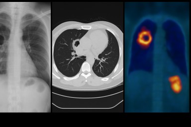
Imaging — especially chest X-ray and high-resolution CT (HRCT) — is one of the most important tools for recognising, diagnosing and monitoring aspergillosis. Because the condition can affect the lungs in very different ways, seeing what is happening inside the chest is essential for both patients and clinicians.
This guide explains why imaging matters, how it is used, and provides links to trusted resources that show what aspergillosis looks like on scans.
Why Imaging Matters in Aspergillosis
Aspergillosis affects the lungs deep within the airway and lung tissue. Many of these changes cannot be detected by a stethoscope or blood tests alone. Imaging helps detect:
-
mucus plugging
-
bronchiectasis (damaged widened airways)
-
cavities (holes) in the lungs
-
fungal balls (aspergillomas)
-
inflammation and consolidation
-
scarring or fibrosis
-
signs of haemoptysis risk
In Allergic Bronchopulmonary Aspergillosis (ABPA), imaging may show mucus impaction or central bronchiectasis.
In Chronic Pulmonary Aspergillosis (CPA), it may show cavities, thickened cavity walls, or a fungus ball.
In invasive aspergillosis, imaging can detect early nodules, the “halo sign”, or rapidly progressing changes.
Understanding these patterns helps clinicians choose the right treatment at the right time, and helps patients make sense of what is happening in their lungs.
Key Online Resources for Aspergillosis Imaging
Radiopaedia – Open Radiology Reference
One of the clearest, most comprehensive imaging resources available.
-
Pulmonary aspergillosis overview:
https://radiopaedia.org/articles/pulmonary-aspergillosis?lang=gb -
Invasive pulmonary aspergillosis:
https://radiopaedia.org/articles/invasive-pulmonary-aspergillosis?lang=gb -
Aspergilloma (fungal ball):
https://radiopaedia.org/articles/aspergilloma?lang=gb
Why it’s useful:
Radiopaedia shows real CT and X-ray examples from multiple patients, helping you understand what radiologists look for and how different forms of aspergillosis appear on imaging.
Peer-Reviewed Pictorial Reviews
These academic reviews provide side-by-side CT examples of the full spectrum of disease, written in an accessible style and extremely useful for both patients and clinicians.
-
Imaging Spectrum in Chronic Pulmonary Aspergillosis (Garg et al., 2022)
https://pmc.ncbi.nlm.nih.gov/articles/PMC9833062/ -
Pictorial Essay: Forms of Pulmonary Aspergillosis (Tamkevičiūtė et al., 2024)
(abstract) https://www.ejradiology.com/article/S0720-048X%2824%2900006-8/abstract
These explain in pictures:
-
how cavities form
-
how aspergillomas look
-
early vs late changes
-
how ABPA patterns differ from CPA
-
what stable vs progressive disease looks like
They are especially helpful for GPs, respiratory trainees, and nurses learning to interpret fungal disease imaging.
Classic RSNA Radiographics Review (Franquet et al.)
A foundational article describing radiologic patterns in allergic, chronic and invasive aspergillosis.
Although technical, this remains a reference standard for understanding how fungal disease presents on CT.
National Aspergillosis Centre (UK) – Patient-Friendly Information
Clear explanations of why imaging is needed, how CT is used, and what typical findings mean.
-
Chronic Pulmonary Aspergillosis (CPA):
https://mft.nhs.uk/wythenshawe/services/infectious-diseases/national-aspergillosis-centre/about-aspergillosis/chronic-pulmonary-aspergillosis-cpa/
These pages are ideal for newly diagnosed patients, people preparing for CT scans, and clinicians who want a quick overview.
Asthma + Lung UK – General Aspergillosis Overview
Patient-friendly explanations of the different types of aspergillosis, diagnosis and treatment.
Includes mention of imaging when relevant.
How Imaging Guides Clinical Decisions
1. Confirming the Diagnosis
Different forms of aspergillosis look different on scans:
-
ABPA may show “finger-in-glove” mucus plugging or central bronchiectasis.
-
CPA requires a cavity seen on imaging for >3 months.
-
Aspergilloma is a fungal ball sitting within a cavity.
-
Invasive disease shows nodules, halo signs, or rapidly evolving infiltrates.
Without CT imaging, these distinctions cannot reliably be made.
2. Checking for Complications
Imaging detects:
-
new cavities
-
cavity wall thickening
-
risk of haemoptysis (bleeding)
-
pleural thickening
-
fibrosis
-
co-existing infections
These findings often trigger urgent or proactive treatment modifications.
3. Monitoring Progression and Treatment Response
Symptoms alone are not enough. Imaging shows whether:
-
disease is stable or progressing
-
antifungal treatment is working
-
inflammation is reducing
-
mucus plugging is clearing
-
new areas are becoming involved
This is why people with CPA or ABPA may have repeat CT scans, usually every 6–12 months or when symptoms worsen.
Using These Resources Safely
These resources are not for self-diagnosis. Instead, they help:
-
patients understand their own scan reports
-
GPs recognise when to refer to a specialist
-
hospital teams spot aspergillosis in complex or unclear cases
-
nurses and allied health professionals visualise lung changes
-
clinicians communicate findings more clearly
Imaging must always be interpreted by a trained radiologist or specialist team, but these materials help demystify the process.
Summary
Imaging is central to the diagnosis and management of aspergillosis. Whether you are a patient trying to understand your scans, a GP seeing a complex chest X-ray, or a hospital clinician assessing breathlessness, these imaging resources provide clear, trustworthy examples.
By learning what different patterns look like — mucus plugging, cavities, aspergillomas, fibrosis or invasive changes — non-specialists can make more confident decisions, and patients can better understand their condition.

Why do some people cough up long, tube-shaped pieces of mucus?
In several chronic lung conditions, the airways can become inflamed and produce thick mucus.
When this mucus sits in the bronchial tubes, it can sometimes harden into a cast shaped exactly like the airway.
People often describe these casts as:
-
long, ribbon-like or “snakeskin” pieces
-
rubbery or stretchy
-
white, yellow, or green
-
shaped like the inside of a tube
Coughing one up can feel dramatic but is usually a sign that your lungs are finally able to clear a blockage.
What does it mean if a cast has black flecks or dark spots?
This can look alarming, but several common, mostly harmless explanations exist.
1. Old or dried blood
Tiny amounts of bleeding from irritated airways can dry and turn:
red → brown → black
This often appears as tiny black dots or threads.
2. Inhaled particles
Dust, soot, pollution, or smoke can get trapped in mucus deeper in the lungs and show up as dark specks.
3. Debris from infection or inflammation
Long-standing inflammation can cause:
-
darkened mucus fragments
-
tiny bits of fungal, bacterial or biofilm material
-
oxidised (darkened) mucus layers
These often look like pepper-like flecks and are not dangerous on their own.
4. Oxidation or ageing of thick mucus
When mucus sits for a long time before it is coughed out, it can become darker in spots.
When this is usually not worrying
Black flecks are often harmless when:
-
the amount is small
-
the colour change is occasional
-
you feel better after coughing the cast out
-
there is no new increase in blood, fever, or breathlessness
-
this fits your usual pattern of mucus plugging
Most people with chronic airway disease experience occasional colour changes in mucus.
When to mention it to your doctor
You should let your team know if:
-
black flecks keep appearing repeatedly
-
you cough up more blood than usual
-
your breathing worsens suddenly
-
your sputum smells different
-
you have fever or chest pain
-
casts become bigger, more frequent, or harder to clear
These changes do not always mean something serious, but they are worth checking.
Why do casts form in the first place?
Conditions that can cause airway casts include:
-
Bronchiectasis
-
ABPA (Allergic Bronchopulmonary Aspergillosis)
-
Severe or eosinophilic asthma
-
Chronic infections, including fungal or bacterial
-
COPD with mucus hypersecretion
Inflammation makes mucus thicker, and narrowed airways make it harder to clear.
Over time, mucus can mould itself into the shape of the airway — becoming a cast.
What to do if you cough one up
-
Stay calm — this often brings relief.
-
Take note of its colour and size.
-
Hydrate well to thin mucus.
-
Continue your usual airway-clearance technique (physio, nebulisers, saline, etc.)
-
Let your team know if it is unusual for you.
Final reassurance
Coughing up a long, tube-like piece of mucus can feel shocking, but in most cases it simply means your lungs are clearing a blocked area.
Black flecks are usually:
-
old blood
-
trapped dust or soot
-
dried mucus debris
Most of the time, these findings are not dangerous, but they can give useful clues about airway inflammation.

🌡️ Understanding Body Temperature in Aspergillosis: Why Your Fever May Look Different
Many people living with aspergillosis—including allergic bronchopulmonary aspergillosis (ABPA), chronic pulmonary aspergillosis (CPA), severe asthma with fungal sensitisation (SAFS) and Aspergillus bronchitis—notice that their body temperature behaves differently from what doctors call “normal.”
This is especially common in people who are:
-
On long-term steroids
-
Tapering steroids
-
Living with adrenal insufficiency
-
Older adults
-
On biologics
-
Managing chronic lung disease
This guide explains why your temperature may run lower, why fevers can appear smaller or absent, and how to safely manage this.
🔶 1. Many aspergillosis patients have a lower baseline temperature
Although “37.0°C” is often quoted, most patients actually sit anywhere between 35.5–36.5°C.
Reasons include:
✔ Long-term steroids
Prednisolone, methylprednisolone, hydrocortisone, and even high-dose inhaled steroids can blunt the immune response and lower your resting temperature.
✔ Adrenal insufficiency
If your adrenal glands are suppressed, your body’s ability to raise temperature is reduced.
You may get no fever at all, even with infections.
✔ Chronic lung disease
Living with ABPA, CPA or bronchiectasis can change how your body regulates heat.
✔ Biologic treatments
Some biologics influence inflammatory signalling and may soften fever responses.
✔ Age
Older adults naturally have:
-
Lower metabolism
-
Lower baseline temperature
-
Reduced ability to generate fever (“immune senescence”)
Many older aspergillosis patients sit around 35.7–36.2°C when completely well.
🔶 2. Fever is a rise from your normal — not a single number
For someone with a naturally low temperature, a fever may look very different.
A useful rule:
A fever = a rise of 1°C above your personal baseline,
even if the thermometer is below 38°C.
Example
-
Your baseline = 35.8°C
-
Your fever may begin at 36.8–37.0°C
You may feel shivery, hot, exhausted or “flu-ish” long before hitting 38°C.
🔶 3. Why fevers are often “muted” in aspergillosis
✔ Steroids
Reduce the body’s ability to trigger a strong fever.
✔ Adrenal insufficiency
Greatly reduces your ability to raise temperature; infections may show as fatigue, dizziness, nausea or sudden weakness instead.
✔ Age
Older adults may have:
-
No fever
-
A tiny rise
-
Confusion or breathlessness as the only sign of infection
✔ Chronic disease
Your temperature regulation system may simply behave differently because of long-term inflammation.
🔶 4. What YOU can do to manage this safely
✔ Know your personal baseline
Measure your temperature twice daily for 5–7 days when well.
Record the average — this is your true normal.
✔ Treat a 1°C rise as your own fever
Don’t wait for the thermometer to reach 38°C.
✔ Watch symptoms more than the number
Seek medical advice if you notice:
-
Feeling feverish or shivery
-
Breathing worsening
-
New chest or flank pain
-
Sudden exhaustion
-
Increased heart rate
-
Confusion, dizziness or “not right”
-
New cough or change in sputum
These can indicate infection even without a high temperature.
✔ Keep a symptom + temperature chart
Especially if you:
-
Are on steroids
-
Have adrenal insufficiency
-
Are tapering
-
Are on biologics
-
Have recurrent infections
Even simple notes help clinicians hugely.
✔ Tell every clinician your temperature baseline
Not all doctors will know your usual pattern, so tell them:
“My normal temperature is around X°C.
I don’t get high fevers because of chronic illness/steroids/adrenal suppression.
A small rise is significant for me.”
This is important in GP appointments, A&E, respiratory clinics and hospital admissions.
🔶 5. Extra precautions if you have adrenal insufficiency
People with steroid-induced adrenal suppression must be especially careful:
-
A small temperature rise + feeling unwell may mean you need stress-dose steroids
-
Vomiting, dizziness, intense fatigue or confusion are warning signs
-
Always follow your adrenal emergency plan
-
Always carry your Steroid Emergency Card and hydrocortisone emergency injection if prescribed
🔶 6. Do doctors understand this?
Most clinicians understand the general rules:
-
Older adults often do not mount high fevers
-
Steroids blunt fever
-
Adrenal insufficiency changes the febrile response
-
Infection may present atypically
However, few clinicians know your personal baseline unless you tell them.
Sharing your own numbers helps them interpret your symptoms safely and accurately.
🟩 Summary for Aspergillosis Patients
-
Many people with aspergillosis have a naturally lower temperature.
-
Steroids, adrenal insufficiency and age can all reduce your ability to produce a fever.
-
A rise of 1°C above YOUR normal may be your fever.
-
Focus on overall symptoms, not just the thermometer.
-
Tell every clinician your baseline temperature.
-
Take extra care if you have adrenal insufficiency.