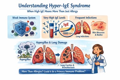
Hyper-IgE syndrome
A patient-friendly guide (and why it matters if you have aspergillosis)
It is not the same as having lots of allergies, even though it can look very similar at first.
What is IgE, and why does it matter?
IgE is usually involved in allergies and asthma.
In Hyper-IgE syndrome:
-
IgE levels are extremely high (often many thousands)
-
But the immune system is unbalanced
-
This makes infections—especially in the lungs and skin—harder to control
So IgE is high, but protection is weak.
How might Hyper-IgE syndrome affect everyday life?
Not everyone has the same symptoms, but common features include:
Lung and chest problems
-
Repeated chest infections (often from a young age)
-
Ongoing cough, breathlessness and mucus
-
Lung damage such as bronchiectasis
-
Lung cavities that can later become infected by moulds such as Aspergillus
Skin and infection problems
-
Long-standing eczema or very sensitive skin
-
Recurrent skin infections or boils
-
Infections that keep coming back or take a long time to clear
Other clues (in some people)
-
Frequent infections in childhood
-
Bone or joint problems
-
Dental issues (for example baby teeth not falling out on time)
Why is this important for people with aspergillosis?
For many people, Aspergillus causes allergy or irritation.
In Hyper-IgE syndrome:
-
The immune system struggles to control moulds
-
Aspergillus can behave more like a true infection, not just an allergy
-
Lung damage can happen more easily and progress faster
This means doctors may need to:
-
Monitor lungs more closely
-
Treat fungal disease earlier and for longer
-
Be cautious with repeated or long-term steroid use
Specialist centres such as the National Aspergillosis Centre are often involved when aspergillosis and immune problems overlap.
Isn’t this just severe allergy or ABPA?
Hyper-IgE syndrome can look similar to:
-
Severe allergic asthma
-
Allergic Bronchopulmonary Aspergillosis (ABPA)
The key difference is that in Hyper-IgE syndrome:
-
The immune system itself is faulty
-
High IgE is part of a wider immune problem
-
Treating allergy alone may not be enough
Some people are treated for asthma or ABPA for years before this possibility is considered.
How is Hyper-IgE syndrome treated?
There is no single cure, but good treatment can make a big difference. The aim is to prevent infections, protect the lungs, and reduce symptoms.
1. Preventing infections (most important)
Because the immune system does not fight germs normally:
-
Some people take regular low-dose antibiotics
-
Others use antibiotics early and promptly when infections start
For people with aspergillosis:
-
Antifungal medicines may be needed
-
Monitoring is usually closer and longer-term
2. Protecting the lungs
Many people develop bronchiectasis or lung damage, so care often includes:
-
Airway clearance physiotherapy
-
Saline nebulisers to help clear mucus
-
Regular sputum tests
-
Early treatment of flare-ups
The goal is to stop the cycle of:
infection → inflammation → permanent lung damage
3. Managing inflammation and allergy (carefully)
People may also have asthma-like symptoms, eczema and multiple allergies.
-
Steroids can help symptoms, but long-term or frequent use can increase infection risk
-
Doctors usually try to keep steroid doses as low as possible
Biologic treatments (such as anti-IgE medicines):
-
May help some people
-
Do not fix the immune problem
-
Are considered on an individual basis, usually in specialist centres
4. Skin care
-
Regular moisturising
-
Prompt treatment of infected eczema
-
Good skin care helps reduce infection risk
How is Hyper-IgE syndrome diagnosed?
Diagnosis usually involves:
-
A detailed review of your medical history (often including childhood infections)
-
Blood tests of immune function
-
Referral to an immunology specialist
-
Sometimes genetic testing
Does having high IgE mean I definitely have this?
No.
Hyper-IgE syndrome is rare.
But it may be worth asking about if:
-
Your IgE has always been extremely high
-
You’ve had repeated infections for many years
-
You have bronchiectasis without a clear cause
-
Aspergillosis seems unusually persistent or severe
-
Standard asthma or allergy treatments don’t fully explain your symptoms
Key message
Very high IgE does not always mean “just allergy.”
In a small number of people, it reflects a deeper immune problem that changes how aspergillosis behaves and how it should be treated.
If your illness doesn’t quite fit the usual labels, it is reasonable to ask whether an immunology review would help.

Sinusitis in Patients with ABPA
When to suspect it, when to investigate, and when to refer
Why this matters
Patients with allergic bronchopulmonary aspergillosis (ABPA) are usually managed as having a lung disease. Diagnosis, monitoring, and treatment focus appropriately on the chest, immunology, and asthma control.
However, ABPA occurs within a single continuous airway, extending from the nose and sinuses to the lungs. Disease in the upper airway can coexist with, exacerbate, or complicate lower airway inflammation — yet sinus disease is not routinely assessed in ABPA care pathways.
This article outlines:
-
What is known about sinus disease in this context
-
Which symptoms should raise suspicion
-
When investigation or ENT referral should be considered
-
What GPs and non-specialists can reasonably do
The united airway: a brief reminder
The upper and lower airways share:
-
Type 2 (eosinophilic) inflammation
-
Immunoglobulin E–mediated immune responses
-
Common triggers, including allergens and fungi
Chronic rhinosinusitis is common in asthma and severe asthma, and treatment of sinus disease can improve lower airway outcomes in some patients.
ABPA sits within this same inflammatory spectrum, even though its management is lung-centred.
Sinus disease in ABPA: what is (and isn’t) known
What we know
-
Chronic rhinosinusitis is common in patients with asthma and severe asthma
-
Sinus disease may be symptomatic or relatively silent
-
ABPA guidelines do not mandate routine ENT review or sinus imaging
-
ENT involvement, therefore, varies widely between centres
What we do not know
-
Whether routine ENT assessment improves ABPA outcomes
-
Which ABPA patients benefit most from sinus intervention
-
The optimal timing for ENT referral in ABPA
As a result, clinical judgement remains central.
Symptoms that should prompt consideration of sinus disease
Sinusitis in ABPA patients does not always present with classic “blocked nose and facial pain”.
Key symptoms include:
Common but often overlooked
-
Persistent post-nasal drip
-
Foul, bitter, metallic, or “infected” taste in the mouth
-
Throat clearing, chronic cough
-
Thick or sticky mucus sensation
-
Symptoms are worse on waking or lying flat
More typical sinonasal features
-
Nasal blockage or congestion
-
Facial pressure or fullness
-
Reduced or altered sense of smell
-
Nasal crusting or discharge
Contextual clues
-
Poor durability of response to steroids or antifungals
-
Recurrent “flares” without clear chest triggers
-
Coexisting severe asthma or nasal polyps
-
Symptoms are worse in damp or mould-affected housing
A persistent foul taste in the mouth is a recognised symptom of chronic sinus disease, usually due to post-nasal drainage of inflamed secretions.
Damp homes and sinus disease
Living in damp or mould-affected environments is associated with:
-
Higher rates of chronic rhinosinusitis
-
Upper airway irritation and inflammation
-
Allergic sensitisation to fungal spores
In most cases, this results in inflammatory or allergic sinusitis, not invasive fungal infection.
Fungal involvement may act as an immune trigger, even when not labelled as “fungal sinusitis”.
Fungal sinusitis: rare vs under-recognised
It is important to distinguish between entities:
| Type | Frequency | Key point |
|---|---|---|
| Invasive fungal sinusitis | Rare | Usually immunocompromised; dramatic presentation |
| Fungal ball (mycetoma) | Uncommon | Usually obvious on CT |
| Allergic fungal rhinosinusitis | Likely under-recognised | Requires active suspicion |
Allergic fungal rhinosinusitis overlaps biologically with ABPA:
-
IgE-mediated
-
Eosinophilic inflammation
-
Thick allergic mucin
It is not routinely sought, so it may be under-diagnosed in at-risk groups.
What GPs and non-specialists can reasonably do
1. Take upper airway symptoms seriously
Especially in ABPA or severe asthma patients with:
-
Persistent post-nasal symptoms
-
Foul taste
-
Recurrent unexplained deterioration
2. Examine the nose and throat
-
Look for polyps, discharge, and crusting
-
Note mouth breathing or altered voice quality
-
Check dentition (to exclude dental causes)
3. Consider imaging when symptoms persist
-
CT sinuses (not plain X-ray) is the imaging of choice
-
Particularly appropriate if symptoms last >8–12 weeks or recur
4. Refer to ENT when:
-
Symptoms are persistent or progressive
-
CT shows significant sinus disease
-
There is a poor response to standard medical therapy
-
There is diagnostic uncertainty
Referral does not imply surgery — ENT input may be diagnostic or medical.
What this article is not saying
-
It does not suggest that all ABPA patients need an ENT referral
-
It does not claim that sinus treatment improves ABPA outcomes
-
It does not override existing guidelines
It does suggest that earlier consideration of the upper airway is reasonable in selected patients.
Key take-home points for clinicians
-
The airway functions as a single inflammatory system
-
Sinus disease may be subtle, under-reported, or atypical
-
A foul taste in the mouth is a meaningful symptom
-
Damp or mould exposure increases sinus disease risk
-
ENT referral is appropriate when symptoms persist or recur
-
Evidence gaps remain — but clinical vigilance is justified
In summary
ABPA is managed as a lung disease, but patients live with a whole airway.
Recognising when sinus disease may be contributing can help explain persistent symptoms and guide appropriate referral — without over-investigation or over-treatment.

ABPA and Work: What a Patient Poll Tells Us About Employment, Health, and Real-World Impact
An article for patients, GPs, and non-specialist healthcare professionals
Allergic bronchopulmonary aspergillosis (ABPA) is often discussed in terms of lung function, immunology, and imaging. Far less often do we talk about its impact on everyday life, particularly on a person’s ability to work.
A poll run within the National Aspergillosis Centre patient community asked a simple but powerful question:
Who is still able to work while living with ABPA – and who has had to stop or retire?
The responses provide an important insight into the functional and socioeconomic burden of ABPA.
Key findings from the poll (patient-reported)
-
Working full time: 17%
-
Working part time (days or hours): 18% combined
-
Not working: 30%
-
Retirement age: 21%
-
Retired early for health reasons: 12%
-
Currently on sick leave / full-time carer / pre-diagnosis: small but notable groups
Even allowing for the informal nature of a social media poll, the overall pattern is clear.
What this tells us
1. Sustained full-time work is uncommon in ABPA
Fewer than one in five respondents were able to work full time. Even among those still working, many described reduced hours, flexible arrangements, or fragile employment dependent on day-to-day health.
ABPA is often incompatible with predictable, high-demand working patterns.
2. ABPA frequently leads to work loss or early retirement
A substantial proportion of respondents were either:
-
No longer working at all, or
-
Retired earlier than planned specifically because of health
This is particularly striking given that ABPA often affects people during their working years and may coexist with asthma, bronchiectasis, or long-term steroid use.
3. “Retirement age” can hide health-forced exit
Some respondents selected “retirement age,” but accompanying comments revealed that many:
-
Left work earlier than expected
-
Changed careers or reduced responsibilities years before retirement
-
Worked through ill health until they no longer could
This matters when interpreting employment statistics: health-driven work loss may be underestimated.
4. Unpaid work and instability are often overlooked
The poll also highlighted:
-
People currently on prolonged sick leave
-
Full-time unpaid carers
-
Individuals still awaiting diagnosis but already struggling to work
These groups are frequently invisible in employment data, yet represent significant personal and societal impact.
Why ABPA affects the ability to work
For patients and non-specialists, it is important to understand that work difficulties in ABPA are not simply due to “asthma symptoms.”
Common contributors include:
-
Chronic breathlessness and cough
-
Severe fatigue and post-exertional exhaustion
-
Recurrent chest infections
-
Steroid side-effects (muscle weakness, bone disease, mood changes, diabetes risk)
-
Unpredictable flare-ups requiring rest, antibiotics, or hospital care
-
Cognitive and emotional burden of long-term illness
Together, these make consistent attendance, physical work, and high cognitive load difficult to sustain.
Implications for patients
-
Difficulty working is not a personal failure
-
Many others with ABPA face similar challenges
-
Adjustments, reduced hours, or stopping work altogether may be medically appropriate
-
Asking for support is reasonable and justified
Implications for GPs and non-specialist clinicians
-
Employment status should be considered a key outcome of disease control
-
Fit notes, occupational health input, and benefits documentation are part of holistic care
-
ABPA is a fluctuating condition – patients may cope for periods and then deteriorate
-
Statements such as “lung function is stable” do not always reflect real-world functioning
Understanding the work impact helps clinicians better support patients in consultations, reports, and advocacy.
Implications for systems and policy
This poll reinforces that ABPA carries a significant socioeconomic burden, including:
-
Reduced workforce participation
-
Early retirement
-
Increased reliance on health and social support systems
Any assessment of disability, employment capability, or long-term planning must take into account:
-
Variability over time
-
Treatment burden
-
Side-effects of necessary medications
In summary
This patient poll sends a consistent message:
ABPA commonly limits the ability to work, often leading to reduced hours, unstable employment, or early exit from the workforce.
For patients, this experience is shared and valid.
For clinicians, it is a reminder that ABPA is not just a radiological or immunological diagnosis, but a life-limiting condition with real-world consequences.

Why do some people cough up long, tube-shaped pieces of mucus?
In several chronic lung conditions, the airways can become inflamed and produce thick mucus.
When this mucus sits in the bronchial tubes, it can sometimes harden into a cast shaped exactly like the airway.
People often describe these casts as:
-
long, ribbon-like or “snakeskin” pieces
-
rubbery or stretchy
-
white, yellow, or green
-
shaped like the inside of a tube
Coughing one up can feel dramatic but is usually a sign that your lungs are finally able to clear a blockage.
What does it mean if a cast has black flecks or dark spots?
This can look alarming, but several common, mostly harmless explanations exist.
1. Old or dried blood
Tiny amounts of bleeding from irritated airways can dry and turn:
red → brown → black
This often appears as tiny black dots or threads.
2. Inhaled particles
Dust, soot, pollution, or smoke can get trapped in mucus deeper in the lungs and show up as dark specks.
3. Debris from infection or inflammation
Long-standing inflammation can cause:
-
darkened mucus fragments
-
tiny bits of fungal, bacterial or biofilm material
-
oxidised (darkened) mucus layers
These often look like pepper-like flecks and are not dangerous on their own.
4. Oxidation or ageing of thick mucus
When mucus sits for a long time before it is coughed out, it can become darker in spots.
When this is usually not worrying
Black flecks are often harmless when:
-
the amount is small
-
the colour change is occasional
-
you feel better after coughing the cast out
-
there is no new increase in blood, fever, or breathlessness
-
this fits your usual pattern of mucus plugging
Most people with chronic airway disease experience occasional colour changes in mucus.
When to mention it to your doctor
You should let your team know if:
-
black flecks keep appearing repeatedly
-
you cough up more blood than usual
-
your breathing worsens suddenly
-
your sputum smells different
-
you have fever or chest pain
-
casts become bigger, more frequent, or harder to clear
These changes do not always mean something serious, but they are worth checking.
Why do casts form in the first place?
Conditions that can cause airway casts include:
-
Bronchiectasis
-
ABPA (Allergic Bronchopulmonary Aspergillosis)
-
Severe or eosinophilic asthma
-
Chronic infections, including fungal or bacterial
-
COPD with mucus hypersecretion
Inflammation makes mucus thicker, and narrowed airways make it harder to clear.
Over time, mucus can mould itself into the shape of the airway — becoming a cast.
What to do if you cough one up
-
Stay calm — this often brings relief.
-
Take note of its colour and size.
-
Hydrate well to thin mucus.
-
Continue your usual airway-clearance technique (physio, nebulisers, saline, etc.)
-
Let your team know if it is unusual for you.
Final reassurance
Coughing up a long, tube-like piece of mucus can feel shocking, but in most cases it simply means your lungs are clearing a blocked area.
Black flecks are usually:
-
old blood
-
trapped dust or soot
-
dried mucus debris
Most of the time, these findings are not dangerous, but they can give useful clues about airway inflammation.

⭐ Severe Asthma with Fungal Sensitisation (SAFS): The Hidden Burden Behind Difficult Asthma
Estimated prevalence: 15–30% of severe asthma patients show fungal sensitisation.
Severe Asthma with Fungal Sensitisation (SAFS) describes a group of patients with severe asthma who show sensitisation (allergy) to Aspergillus or other environmental moulds but do not meet criteria for ABPA. These patients often experience persistent inflammation, breathlessness, mucus production, and exacerbations that are not adequately controlled by standard asthma therapies.
Although SAFS is common in severe asthma clinics, it remains poorly recognised, frequently mislabelled, and rarely discussed in routine practice. Yet identifying SAFS is crucial because it opens the door to specific interventions — including antifungals or targeted biologics — that can improve symptoms and reduce hospital admissions.
⭐ How Common Is SAFS?
SAFS is more common than ABPA and CPA combined in asthma services.
| Population | Estimated prevalence |
|---|---|
| Moderate asthma | ~5% |
| Severe asthma | 15–30% |
| Patients with frequent exacerbations | up to 40% |
| ABPA-negative patients with mucus plugging | high likelihood |
Across the UK, this represents tens of thousands of people.
⭐ Why SAFS Is Missed
1. The diagnosis is not widely understood
Unlike ABPA or CPA, SAFS lacks:
-
universally agreed diagnostic criteria
-
clear imaging features
-
a single confirmatory test
This leads to variability in thinking and detection.
2. Symptoms mimic uncontrolled asthma
SAFS patients typically experience:
-
severe breathlessness
-
wheeze
-
mucus production
-
airway plugging
-
poor response to inhalers
-
frequent steroid courses
These appear indistinguishable from “difficult” or “type 2–high” asthma.
3. IgE and eosinophils may be normal
Unlike ABPA:
-
total IgE may be modest
-
Aspergillus IgE may be borderline
-
eosinophils may fluctuate, especially with steroids or biologics
Clinicians are often looking for very high IgE levels — but SAFS patients usually don’t show them.
4. Sputum and CT scans appear non-specific
Typical imaging:
-
mucus plugging
-
small-airway thickening
-
variable, patchy inflammation
-
bronchiectasis may or may not be present
Radiologists often report these changes as:
-
“consistent with asthma”
-
“post-infective”
-
“non-specific inflammatory pattern”
5. The fungal link is overlooked
Many clinicians are unfamiliar with:
-
the role of mould exposure
-
sensitisation thresholds
-
the overlap between environmental allergy and airway disease
-
when antifungals are appropriate
This leads to delays in recognising fungal-driven asthma.
⭐ Who Is at Highest Risk?
1. Severe asthma patients unresponsive to maximal inhaled treatment
Particularly those with:
-
frequent exacerbations
-
nocturnal symptoms
-
long-term steroid use
-
persistently low lung function
-
mucus plugging events
2. Patients sensitised to Aspergillus or multiple moulds
Positive skin tests or specific IgE indicate airway allergy that can drive symptoms.
3. Patients with damp or mould exposure at home or work
An important environmental factor often overlooked.
4. ABPA-negative asthma patients with mucus plugging
A large proportion of these patients fit the SAFS profile.
5. Those with co-existing bronchiectasis
Bronchiectasis amplifies the inflammatory response to fungal exposure.
⭐ Specialties That Need Greater Awareness
-
Severe asthma services & biologics clinics
(primary diagnostic opportunity) -
General respiratory clinics
-
Primary care & urgent care
(patients seen frequently with “persistent asthma symptoms”) -
Radiology
(important for identifying mucus plugging) -
Allergy/Immunology
(mould sensitisation is central to diagnosis) -
Environmental health teams
(exposure to mould and dampness often perpetuates symptoms)
The National Aspergillosis Centre can provide specialist input when diagnosis is unclear or response to treatment is suboptimal.
⭐ Red Flags Suggesting SAFS
1. Severe asthma poorly controlled despite maximal inhalers
Including biologics (omalizumab, mepolizumab, benralizumab, dupilumab, tezepelumab).
2. Sensitisation to Aspergillus fumigatus or multiple moulds
3. Repeated mucus plugging episodes
(or “sticky mucus” symptoms)
4. More than 2–3 steroid-treated exacerbations per year
5. Asthma + bronchiectasis
Even mild bronchiectasis increases fungal risk.
6. Symptoms triggered by damp/mould exposure
7. Persistent airway inflammation despite correct inhaler technique
⭐ Misdiagnoses That Delay Recognition
-
“Difficult asthma”
-
“Brittle asthma”
-
“Post-viral inflammation”
-
“Poor adherence to inhalers”
-
“Asthma–COPD overlap”
-
“Psychogenic dyspnoea”
-
“Recurrent chest infections”
SAFS is a diagnosis hiding in these labels.
⭐ The Cost of Missed SAFS Diagnosis
For patients:
-
persistent symptoms
-
steroid dependence
-
increased risk of ABPA
-
progressive airway damage
-
hospital admissions
-
poor quality of life
-
possible career and lifestyle impact
For healthcare systems:
-
repeated A&E visits
-
asthma admissions
-
high biologic usage without adequate response
-
unnecessary antibiotics
-
escalating steroid toxicity
-
missed environmental interventions
⭐ Conclusion
SAFS is one of the most common — yet least recognised — fungal-related lung conditions. Although it lacks the dramatic imaging changes of ABPA or CPA, its impact on patients is profound.
Recognising mould sensitisation in severe asthma, understanding the role of fungal allergens, and considering targeted therapies can transform disease control. For complex cases or when the diagnosis is uncertain, referral to the National Aspergillosis Centre is recommended.
Early identification and appropriate treatment reduce steroid use, exacerbations, and long-term airway damage.

⭐ Aspergillus Bronchitis: The Overlooked Condition Hiding in Plain Sight
Estimated prevalence 1–2% in bronchiectasis and chronic airway disease clinics.
Aspergillus Bronchitis (AB) is a chronic, symptomatic infection of the airways caused by Aspergillus species in people with underlying lung disease. It sits between simple colonisation and chronic pulmonary aspergillosis (CPA), and is frequently overlooked or mislabelled as “recurrent infection,” “post-viral symptoms,” or uncontrolled bronchiectasis.
Unlike CPA, Aspergillus Bronchitis does not require cavities or major structural destruction — which makes it both easier to miss and surprisingly common among people with chronic airway disease.
When recognised and treated (usually with antifungal therapy for several months), symptoms often improve significantly. But because awareness remains low, most patients cycle through unnecessary antibiotics, repeated exacerbations, and worsening airway disease before the real cause is identified.
⭐ What Exactly Is Aspergillus Bronchitis?
Aspergillus Bronchitis is defined by:
-
chronic productive cough
-
sputum growing Aspergillus species repeatedly
-
airway inflammation
-
symptoms lasting over 3 months
-
underlying airway disease (bronchiectasis, CF, COPD, prior TB, ABPA)
-
response to antifungal therapy
Unlike ABPA:
-
there is no allergic response,
-
IgE is usually normal,
-
eosinophils are normal or mildly elevated.
Unlike CPA:
-
there are no cavities on imaging,
-
IgG may be normal or only slightly elevated,
-
disease is confined to the airways, not lung tissue.
This places AB in a “grey zone” — often invisible unless specifically looked for.
⭐ Why Aspergillus Bronchitis Is Missed
1. Symptoms mimic common chronic airway disease
Typical AB symptoms include:
-
daily productive cough
-
worsening sputum thickness
-
breathlessness
-
fatigue
-
repeated “chest infections”
-
slow-to-clear mucus
-
crackles or wheeze
These resemble:
-
bronchiectasis exacerbations
-
COPD flare-ups
-
chronic infection with Pseudomonas or NTM
-
post-viral cough
-
uncontrolled asthma
Without fungal awareness, clinicians default to bacterial explanations.
2. Sputum grows multiple organisms — Aspergillus is dismissed
In bronchiectasis, sputum frequently grows:
-
Haemophilus
-
Pseudomonas
-
Staphylococcus
-
Streptococcus
-
NTM
When Aspergillus appears, it’s often labelled:
-
“colonisation”
-
“contaminant”
-
“not clinically relevant”
But repeated isolation with persistent symptoms is highly suggestive of AB.
3. IgE/IgG results may be normal
Many clinicians expect high IgE or IgG to “confirm Aspergillus disease.”
But in Aspergillus Bronchitis:
-
IgE is usually normal
-
IgG can be normal or borderline
This leads to false reassurance.
4. Radiology rarely shows overt features
CT scans in AB may show:
-
mucus plugging
-
mild bronchial wall thickening
-
small nodules
-
progression of bronchiectasis
But they do not show the cavities of CPA or classic features of ABPA.
Therefore radiologists often report scans as “no significant change” or “stable bronchiectasis.”
5. Antibiotics appear to help — temporarily
Patients often improve slightly with:
-
amoxicillin
-
doxycycline
-
macrolides
-
ciprofloxacin
This gives clinicians the impression of bacterial disease, but symptoms soon return.
6. Lack of awareness
Many specialists (even in respiratory clinics) are unaware that Aspergillus Bronchitis:
-
exists as a distinct clinical entity
-
can be disabling
-
responds to antifungals
-
predicts progression to CPA if untreated
This leads to significant diagnostic delay.
⭐ Who Is at Highest Risk?
1. Bronchiectasis
The largest risk group.
Aspergillus Bronchitis may account for 1–2% of all bronchiectasis patients, and up to 5–10% in severe or frequent exacerbator groups.
2. Cystic Fibrosis (CF)
These patients frequently grow Aspergillus but not all have ABPA — some have Aspergillus Bronchitis.
3. COPD and chronic productive cough
Especially those with:
-
frequent mucus plugging
-
repeated “infective exacerbations”
-
progressive sputum production
4. Post-TB airway damage
Chronic airway deformity, scarring, and bronchiectasis from old TB predispose to fungal infection.
5. Post-COVID structural disease
A new and growing risk group, especially after prolonged ICU ventilation.
6. ABPA patients
Some patients develop Aspergillus Bronchitis during steroid-dominated treatment or after stopping antifungals.
⭐ Which Specialities Need Greater Awareness?
-
Respiratory medicine
(especially bronchiectasis clinicians and severe asthma teams) -
Infectious Diseases
(frequent respiratory presentations with chronic airway infection) -
Radiology
(to recognise subtle but progressive airway changes) -
Primary care
(“recurrent chest infection” or “persistent cough” patients) -
Physiotherapy & airway clearance teams
(excessive sputum with fungal elements) -
Cystic Fibrosis services
The National Aspergillosis Centre is the ideal referral destination when diagnosis is uncertain or symptoms persist despite typical management.
⭐ Red Flags Suggesting Aspergillus Bronchitis
1. Chronic (>3 months) productive cough + repeated Aspergillus in sputum
Even 2 positive sputums in the right clinical context should raise suspicion.
2. Bronchiectasis patient not improving on repeated antibiotics
3. Thick, tenacious mucus with black, grey, or brown plugs
4. Worsening CT bronchiectasis or mucus plugging
5. Absence of features typical for ABPA (normal IgE, no fleeting infiltrates)
6. Asthma or COPD patient with new persistent sputum
7. Partial response to antibiotics but rapid relapse
8. Unexplained fatigue and breathlessness in someone with airway disease
⭐ The Cost of Missed Aspergillus Bronchitis
If AB is not recognised early, consequences include:
-
repeated exacerbations
-
accelerating bronchiectasis
-
long-term airway damage
-
chronic inflammation
-
steroid overuse
-
unnecessary antibiotics
-
repeated hospitalisations
-
progression to CPA in some patients
For health systems, missed diagnosis leads to:
-
higher admission rates
-
inappropriate long-term antibiotic use
-
avoidable CT scans and investigations
-
greater long-term burden of CPA
But antifungal therapy — when appropriately used — can offer marked symptom improvement and reduce exacerbation frequency.
⭐ Conclusion
Aspergillus Bronchitis is a distinct, treatable form of chronic airway disease seen in people with bronchiectasis, asthma, COPD, CF, and post-TB lung damage. Yet lack of awareness means many patients are repeatedly misdiagnosed with bacterial infections or unexplained chronic cough.
Recognising red flags, reviewing sputum results carefully, and considering antifungal therapy can dramatically improve outcomes. Early referral to specialist centres such as the National Aspergillosis Centre is recommended for complex cases or uncertain diagnosis.
Early identification prevents airway deterioration — and reduces the likelihood of progression to CPA.

⭐ Allergic Bronchopulmonary Aspergillosis (ABPA): Why Diagnosis Is Missed and Who Needs to Be More Aware
With estimated prevalence of 1–2% in asthma clinics and up to 10% in severe asthma services.
Allergic Bronchopulmonary Aspergillosis (ABPA) is a chronic immune reaction to Aspergillus that affects people with asthma or cystic fibrosis. It causes airway inflammation, mucus plugging, recurrent exacerbations, and bronchiectasis if untreated.
Despite being treatable, ABPA remains heavily underdiagnosed, even in countries with advanced respiratory services. Many people are told for years that they have “difficult asthma” or “recurrent chest infections,” only for ABPA to be diagnosed much later, often with significant lung damage already present.
The UK National Aspergillosis Centre (NAC) provides specialist expertise, yet only a small proportion of expected ABPA cases reach specialist review.
This article explains why ABPA is missed, which patients are at risk, which specialities need to be more alert, and the red flags that should prompt testing or referral.
⭐ How Common Is ABPA?
ABPA is more common than most clinicians realise:
| Population | Estimated prevalence |
|---|---|
| General asthma | 1–2% |
| Severe asthma clinics | 3–10% |
| Cystic fibrosis | 5–15% |
| Bronchiectasis (non-CF) | 1–4% |
Across the UK, this equates to an estimated 15,000–25,000 people living with ABPA — but only a small minority ever receive the correct diagnosis.
⭐ Why ABPA Is Often Missed
1. ABPA looks like “difficult asthma”
Typical symptoms — wheeze, cough, mucus, breathlessness — mimic:
-
severe asthma
-
eosinophilic asthma
-
uncontrolled asthma despite treatment
Patients may be repeatedly stepped up through inhalers, oral steroids, and biologics before ABPA is even considered.
2. Exacerbations are mistaken for infections
Many ABPA flare-ups are treated as:
-
pneumonia
-
viral infection
-
“chest infection”
-
post-viral asthma worsening
Without fungal-specific thinking, the diagnosis is rarely made.
3. IgE and eosinophils fluctuate
IgE is a cornerstone of ABPA diagnosis, but:
-
systemic steroids suppress IgE
-
biologics (benralizumab, mepolizumab, dupilumab) reduce eosinophils
-
flare-ups produce temporary spikes
-
baseline ranges vary between labs
Clinicians often overlook ABPA in patients on biologics because eosinophils are normal — despite the underlying fungal allergy still being active.
4. Radiology findings get mislabelled
ABPA causes:
-
mucus plugging
-
“tram lines” and bronchial thickening
-
fleeting infiltrates
-
upper lobe bronchiectasis
These are often:
-
labelled “infection”
-
attributed to asthma airway remodelling
-
not compared across time
-
missed on CT unless specifically looked for
5. Inconsistent awareness across specialities
Some clinicians are unfamiliar with:
-
ISHAM diagnostic criteria
-
interpreting IgE/IgG results
-
the relationship between asthma and fungal allergy
-
the overlap between ABPA and bronchiectasis
This leads to diagnostic delay or misdiagnosis.
6. ABPA evolves into chronic disease if untreated
Repeated inflammation → mucus plugging → bronchiectasis → fibrosis.
By the time a diagnosis is made, airway damage can be permanent.
⭐ Who Is at Highest Risk?
1. Asthma patients with repeated exacerbations
Especially those who:
-
fail maximal inhaler therapy
-
require multiple steroid courses
-
have sudden, dramatic mucus plugging events
-
experience episodic “flares” with no clear cause
2. Severe asthma clinic patients
Prevalence is up to 10%, especially those with:
-
high IgE
-
eosinophilia
-
sensitisation to multiple allergens
-
steroid dependence
3. Bronchiectasis patients
Bronchiectasis often coexists with ABPA and can worsen flares.
4. Patients with mucus plugging (“finger-in-glove” signs)
These striking CT appearances strongly suggest ABPA but are often misattributed to infection.
5. People with CF (Cystic Fibrosis)
5–15% develop ABPA at some stage.
⭐ Which Specialities Need Greater Awareness?
-
Severe asthma services & biologics clinics
(highest yield group for ABPA detection) -
Respiratory medicine
(diagnosis often falls here but is highly variable) -
General practice
(sees frequent “exacerbations”) -
Emergency departments & acute medical units
(manage acute mucus plugging, chest tightness) -
Paediatric respiratory medicine
(early recognition prevents chronic damage) -
Cystic Fibrosis services
-
Radiology
(fleeting infiltrates and mucus plugging often give the earliest clues)
The National Aspergillosis Centre should be the referral point for complex or uncertain cases.
⭐ Red Flags Suggesting ABPA
1. Asthma with repeated, unexplained exacerbations
Especially if poorly responsive to normal treatment.
2. High total IgE (>500–1000 IU/mL)
Or rising IgE over time.
3. Eosinophilia (unless suppressed by treatment)
4. Positive Aspergillus sensitisation
(Skin prick test or specific IgE)
5. Bronchiectasis, particularly central or upper lobe
6. Fleeting pulmonary infiltrates
7. Mucus plugging on CT (“finger-in-glove” appearance)
8. ABPA flare triggered by stopping antifungals
9. Asthma + Aspergillus in sputum
⭐ The Cost of Missed ABPA Diagnosis
Failure to diagnose ABPA leads to:
-
progressive airway damage
-
permanent bronchiectasis
-
steroid dependence
-
hospital admissions
-
repeated infections
-
respiratory failure in advanced stages
-
reduced quality of life
-
avoidable healthcare expenditure
Delayed diagnosis increases the risk of progression to CPA, a far more serious chronic fungal infection requiring long-term antifungal therapy.
Early recognition, correct treatment, and referral to specialist centres like the National Aspergillosis Centre dramatically improve long-term outcomes.
⭐ Conclusion
ABPA is not rare — especially within severe asthma clinics, bronchiectasis services, and CF units. Yet it remains significantly underdiagnosed because its symptoms mirror those of common respiratory conditions, and because key investigations like IgE, IgG, and CT interpretation are inconsistently used.
A structured approach — recognising red flags, performing appropriate testing, and referring complex cases to the National Aspergillosis Centre — can reduce the burden of avoidable airway damage and improve the lives of thousands of patients.
⭐ Chronic Pulmonary Aspergillosis: Why Diagnosis Is Missed and Who Needs to Be More Aware
With estimated prevalence of 3–4 cases per 100,000 population, and far higher rates in high-risk groups.
Chronic Pulmonary Aspergillosis (CPA) is a slowly progressive fungal lung disease affecting an estimated 3–4 per 100,000 people in the UK, with higher estimates in global settings with greater TB prevalence. Despite this, many clinicians will go through entire careers without confidently recognising it — not because it is extremely rare, but because it almost always hides inside other long-term lung diseases.
The UK is unusual in having a nationally commissioned specialist service — the National Aspergillosis Centre (NAC), based at Wythenshawe Hospital, Manchester — offering funded diagnostics, multidisciplinary review, and long-term antifungal management. But only a fraction of expected CPA cases are ever referred. Most are simply never diagnosed.
This article explains why diagnoses are missed, who is at highest risk, which specialities need to be more alert, and the red flags that should trigger testing or referral.
⭐ How Common Is CPA? The Numbers Behind the Problem
The UK prevalence is estimated at 3–4 per 100,000 people — approximately 2,000–2,500 people with CPA at any given time.
But the risk is far higher in specific groups:
| Risk Group | Estimated CPA prevalence |
|---|---|
| Post-TB lung disease | 6–10% in those with residual cavities |
| Severe COPD (GOLD III–IV) | 1–3% |
| Bronchiectasis | 1–3% |
| NTM disease | 3–10% |
| Sarcoidosis with fibrosis | 1–2% |
| Immunosuppression (steroids/biologics) | Unknown, but rising |
Using these figures, the true UK caseload could exceed 4,000–6,000 individuals, yet NAC receives ~500–1,000 referrals, highlighting a large diagnostic gap.
⭐ Why CPA Is So Often Missed
1. Symptoms mimic common chronic lung diseases
CPA presents with:
-
Persistent cough
-
Breathlessness
-
Fatigue
-
Weight loss
-
Recurrent “chest infections”
-
Haemoptysis
These overlap almost perfectly with:
-
COPD
-
bronchiectasis
-
post-TB changes
-
long COVID
-
NTM infection
-
repeatedly “slow to clear” pneumonia
Because symptoms are non-specific, clinicians rarely think fungal.
2. Interpretation of imaging is inconsistent
CPA shows:
-
one or more cavities
-
pleural thickening
-
nodules
-
progressive changes over months
-
fungal balls
Common reporting pitfalls:
-
labelled “post-infective scarring”
-
misinterpreted as malignancy
-
seen but not compared longitudinally
-
incidental CT findings not acted upon
Radiology is one of the biggest missed opportunities for early detection.
3. IgG testing is not routinely requested
Aspergillus IgG is the key diagnostic biomarker — but it is:
-
often confused with IgE
-
not available in some hospitals
-
omitted from workups for recurrent infection
-
unfamiliar to non-respiratory clinicians
Without IgG, CPA is rarely diagnosed.
4. Short-term improvement with antibiotics is misleading
Patients with CPA may temporarily feel better after:
-
broad-spectrum antibiotics
-
steroids
-
physiotherapy
This transient improvement creates false reassurance.
5. CPA spans multiple specialisms — and no one owns it
Diagnosis requires combined expertise across:
-
respiratory medicine
-
infectious diseases
-
radiology
-
microbiology
-
immunology
When no one speciality takes responsibility, patients get lost.
⭐ Which Patients Are at High Risk?
CPA almost always develops on a background of existing lung damage.
1. Post-TB lung disease (PTLD)
Globally the largest CPA population.
Residual cavities are the strongest predictor.
Specialities needing awareness:
-
TB teams
-
ID physicians
-
Radiologists
-
Community TB nurses
-
Public health TB programmes
2. COPD (especially severe / emphysema)
Millions of people are potentially at risk.
Recurrent infections + bullae/cavities = fertile ground for CPA.
Specialities:
-
COPD clinics
-
Pulmonary rehab
-
Acute medicine (frequent admissions)
3. Bronchiectasis
Damaged airways enable persistent Aspergillus colonisation and inflammation.
Specialities:
-
Bronchiectasis MDTs
-
Severe asthma & NTM clinics
-
Respiratory physiotherapy
4. Sarcoidosis and ILD
Fibrosis and traction bronchiectasis develop cavities over time.
5. Post-COVID or post-influenza structural disease
Emerging risk group, especially in patients with:
-
ventilatory lung injury
-
persistent CT abnormalities
-
chronic steroid exposure
6. Chronic steroid or immunomodulator use
While invasive aspergillosis is linked to profound immunosuppression, CPA often affects those with milder, chronic immune dysfunction:
-
systemic steroids
-
high-dose inhaled steroids
-
biologics affecting eosinophils
-
poorly controlled diabetes
-
chronic kidney disease
-
malnutrition
⭐ Which Specialities Need to Be More Alert?
-
Respiratory Medicine – primary detection, but awareness varies greatly
-
Infectious Diseases – especially post-TB and persistent infection clinics
-
Radiology – key to spotting early changes
-
Primary Care – sees patients repeatedly with “ongoing chest infections”
-
Emergency & acute medicine – haemoptysis presentations
-
Bronchiectasis and NTM services – strong overlap
-
Severe asthma and biologics teams – ABPA → CPA evolution
-
TB clinics – highest prevalence globally, often least recognised
The National Aspergillosis Centre should be the referral point for any complex or uncertain case.
⭐ Red Flags: When to Suspect CPA
1. Cavities on CT (thin-, thick-walled, evolving, or multiple)
Especially with pleural thickening.
2. Haemoptysis
CPA is one of the most common causes of haemoptysis in people with cavities.
3. Symptoms lasting >3 months
Chronic cough, fatigue, weight loss, breathlessness.
4. “Recurrent infections” that never fully resolve
5. Post-TB patient with any new or worsening symptoms
6. Bronchiectasis patient with new cavity or Aspergillus culture
7. High or rising Aspergillus IgG
8. ABPA patient who deteriorates off antifungals
⭐ The Cost of Missed Diagnoses
When CPA is not recognised early, the consequences are severe:
-
irreversible lung damage
-
repeated hospitalisations
-
emergency haemoptysis events
-
prolonged antifungal therapy with more toxicity
-
reduced quality of life
-
avoidable deaths
For systems like the NHS, late diagnosis increases costs:
-
unplanned admissions
-
repeated CT imaging
-
prolonged antibiotics
-
intensive care during haemoptysis
-
complex surgery (lobectomy/pneumonectomy)
Early referral to specialist centres like the National Aspergillosis Centre prevents many of these harms.
⭐ Conclusion
CPA is not rare within the populations most likely to develop it.
Missed diagnoses are common, predictable, and preventable.
By increasing awareness across Respiratory, Infectious Diseases, Radiology, Primary Care, TB services, and severe asthma pathways — and by using simple tools such as Aspergillus IgG and careful CT interpretation — clinicians can dramatically reduce the diagnostic delay that damages lungs, quality of life, and survival.
The UK is fortunate to have the National Aspergillosis Centre as a nationally commissioned referral service. Recognising CPA early and referring appropriately has the power to save lives, reduce system costs, and improve long-term outcomes.

🌿 Biologics when ABPA and CPA overlap: What Patients Need to Know
Understanding how they work, when they’re helpful, and when extra care is needed
Biologic medicines (such as omalizumab, mepolizumab, benralizumab, dupilumab and newer options like tezepelumab) are increasingly used to treat Allergic Bronchopulmonary Aspergillosis (ABPA) and severe asthma. They can be life-changing for some people.
However, their place in Chronic Pulmonary Aspergillosis (CPA) — especially in people who have both ABPA and CPA together — is more complicated and needs careful specialist supervision.
This article explains what we know so far.
🌟 1. ABPA and CPA are different conditions — but some people have both
-
ABPA is mainly an allergic reaction to Aspergillus in the airways.
-
CPA is a chronic fungal infection that causes cavities, scarring, and long-term lung damage.
-
Some people start with ABPA and later develop CPA, or the two conditions overlap.
-
The 2024 international ABPA guidelines now recognise this overlap as real and important.
Because biologics target allergy pathways rather than fungal infection, treatment decisions must look at both sides of the disease.
🌿 2. Biologics in ABPA: the evidence is strong and growing
Biologics can help patients with ABPA or severe asthma by:
-
reducing steroid use
-
improving breathing
-
decreasing mucus plugging
-
lowering flare-ups
-
improving quality of life
Biologics most commonly used in ABPA include:
| Biologic | Target | Notes |
|---|---|---|
| Omalizumab | IgE | Well established, helps many ABPA patients |
| Mepolizumab | IL-5 | Helps eosinophilic inflammation |
| Benralizumab | IL-5Rα | Similar to mepolizumab; long-acting |
| Dupilumab | IL-4Rα | Very promising for allergic disease; growing evidence for ABPA |
| Tezepelumab | TSLP | Very new; limited ABPA data so far |
For many people with ABPA, biologics are safe and effective when monitored.
⚠️ 3. Biologics and CPA: much less evidence
-
CPA is caused by persistent fungal infection and structural lung damage.
-
Biologics do not treat fungal infection, and they do not prevent cavities.
-
In CPA, the mainstay of treatment is still:
-
antifungal medication (usually itraconazole, voriconazole or posaconazole)
-
careful imaging (CT scans)
-
airway clearance
-
sometimes surgery or bronchoscopy
-
There is no strong evidence that biologics help CPA itself.
🔄 4. What about patients who have both ABPA and CPA?
This is where things become more complex.
Biologics may help the allergic part (ABPA), but:
-
they do not treat fungal infection
-
they do not stop fungal cavities from progressing
-
they may reduce inflammation that normally helps the body contain infection
If antifungal treatment is interrupted or not strong enough, fungal activity may increase while the allergic symptoms improve — so regular monitoring is essential.
Specialist centres (like the NAC) now emphasise:
✔️ Continue antifungals if CPA is active
✔️ Watch cavities with regular CT scans
✔️ Monitor Aspergillus IgG/IgE and fungal cultures
✔️ Check whether symptoms are from allergy, infection, or both
✔️ Make joint plans between asthma/airway doctors and mycology specialists
❓ 5. Are some biologics better than others for ABPA/CPA overlap?
There is no official guidance yet, but early observations suggest:
⭐ Most promising for ABPA:
-
Dupilumab seems particularly effective for allergic disease (IgE, mucus, airflow), though still off-label for ABPA.
⭐ Increasing interest:
-
Tezepelumab works outside the eosinophil pathway and may be useful in some asthma types, but research in ABPA is only just starting.
⭐ Useful in selected cases:
-
Anti-IL-5 biologics (mepolizumab, benralizumab) help airway eosinophils but may not help every ABPA patient.
⚠️ Uncertain in CPA:
-
None of the biologics treat fungal infection or cavities directly.
-
Their role in active CPA remains unclear and requires careful oversight.
🧭 6. What this means for patients
If you have ABPA only, biologics may be an excellent option — especially if:
-
steroids cause side-effects
-
your asthma is uncontrolled
-
you have frequent flare-ups
-
your IgE levels are very high
-
mucus plugging or wheezing continues despite treatment
If you have CPA or cavities, treatment needs to be more cautious:
-
antifungal medication usually needs to continue
-
biologics may still help if the allergic component is significant
-
CT scans must be repeated to make sure cavities are not progressing
-
specialists must weigh benefits vs. risk for each patient individually
💬 7. Summary
-
Biologics can be extremely helpful for ABPA.
-
They do not treat CPA, and cannot replace antifungal medicines.
-
In patients with both ABPA and CPA, the approach must be personalised.
-
Dupilumab and (possibly) tezepelumab are emerging biologics with promise, but evidence is still developing.
-
Decisions should always be made with a specialist centre such as the National Aspergillosis Centre (NAC).

Understanding Mucous Casts in Allergic Bronchopulmonary Aspergillosis (ABPA)
People living with Allergic Bronchopulmonary Aspergillosis (ABPA) often notice thick, unusual mucus coming up during a flare. Some of this mucus can look very different from “normal” sputum and may be described as mucous casts. This leaflet explains what they are, why they happen, and what they mean for your ABPA.
⭐ What are mucous casts?
A mucous cast is a thick, sticky plug of mucus that forms inside your airways.
It takes on the exact shape of the airway or branch it was sitting in – a bit like a soft mould of the inside of your lungs.
When coughed up, casts may look:
-
long and tube-shaped
-
soft and rubbery
-
curled or C-shaped
-
occasionally branching, like a twig
-
pale yellow/cream with darker specks
These darker flecks can include dead inflammatory cells, airway debris, and sometimes tiny amounts of fungal material trapped inside.
⭐ Why do they happen in ABPA?
ABPA is not an infection, but an allergic over-reaction to the Aspergillus fungus.
This allergic inflammation causes:
1. Excess mucus production
Your airways create far more mucus than usual.
2. Thicker, stickier mucus
Inflammation changes the chemistry of the mucus, making it harder to clear.
3. Swollen, narrowed airways
This makes it easy for mucus to get stuck and form plugs.
4. Trapped material
Casts can contain:
-
fungal spores
-
inflammatory cells
-
dust or other inhaled particles
-
old blood or tissue debris
All of this can glue together into a cast.
⭐ Are mucous casts harmful?
They are not dangerous on their own, but they can cause problems:
-
Airway blockage → breathlessness, wheeze, sudden tightness
-
Chest infections → trapped mucus is an ideal place for bacteria
-
ABPA flare-ups → casts often appear during periods of high inflammation
-
Reduced airflow on CT scans → seen as “bronchial impaction”
Telling your clinical team when you notice casts helps them judge how active your ABPA is.
⭐ What do mucous casts look like in ABPA?
Patients often describe:
-
“noodles”
-
“worms”
-
“rubbery plugs”
-
“little branches”
-
“specks of brown/black” within pale mucus
These appearances are normal in ABPA and do not mean your lungs are permanently worsening.
⭐ How are mucous casts managed?
1. Airway clearance
This is the most important step. Techniques include:
-
huff-coughing
-
active cycle of breathing
-
nebulised saline (hypertonic or isotonic)
-
flutter/PEP devices (Acapella, Aerobika)
-
chest physiotherapy
These help loosen and move mucus from deeper airways.
2. Medication
Depending on your treatment plan:
-
inhalers (bronchodilator + inhaled steroids)
-
biologics (e.g., mepolizumab, dupilumab, omalizumab)
-
antifungal medication if prescribed as part of your ABPA care
-
oral steroids if medically appropriate
Biologics can reduce the inflammation that causes casts, so many patients notice fewer plugs over time.
3. Monitoring
Your team may keep an eye on:
-
sputum samples
-
IgE levels
-
CT scan changes
-
symptom patterns
⭐ When should I tell my team?
Contact your clinical team if you notice:
-
more frequent mucous casts
-
sudden breathlessness or chest tightness
-
a drop in your usual oxygen saturation
-
fever or signs of infection
-
coughing up blood
-
a change in colour or smell of mucus
⭐ Reassurance
Mucous casts are very common in ABPA.
They can look alarming, but they are simply a sign that your airways are inflamed and producing thick mucus.
Coughing them out is helpful, not harmful.
It allows the affected airway to reopen and can rapidly improve breathing.
✅ Further Reading
For more patient-oriented information, you can visit the AFIT website where the term “casts” is discussed in the context of aspergillosis: Aspergillus.org.uk – search “casts”.