Weekly Aspergillosis Research Update: COPD IPA Criteria, CPA Serology, ICU Galactomannan and Environmental Prevention
Week ending 6 July 2026
Overall summary
This week’s strongest theme is improved recognition and interpretation of Aspergillus disease in high-risk respiratory and immunocompromised patients.
The headline paper proposes COPD-specific diagnostic criteria for invasive pulmonary aspergillosis (IPA), addressing a long-standing gap between classic immunocompromised-host definitions and real-world respiratory practice. Other important papers focus on chronic pulmonary aspergillosis (CPA) serology, bronchoalveolar lavage galactomannan stewardship, endotracheal aspirate galactomannan in ICU patients, and environmental prevention of invasive fungal disease in paediatric cancer care.
Overall, this is a week of cautious progress: better criteria, better test interpretation, and better diagnostic systems — but several findings still require prospective validation.
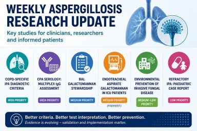
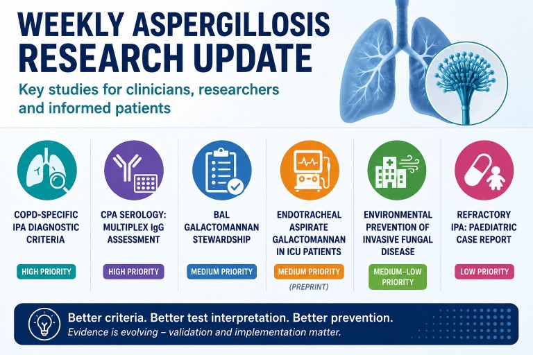
High priority
Diagnostic criteria for invasive pulmonary aspergillosis in COPD patients
Denning DW, Rogers TR, Takazono T, Su X, Lagrou K, White PL, James DA, Bafadhel M, Lopez JB, Bulpa P, Chotirmall SH, et al.
American Journal of Respiratory and Critical Care Medicine. Published 1 July 2026.
DOI: 10.1093/ajrccm/aamag310
PMID: 42384914
This is the likely headline paper of the week. It proposes COPD-specific diagnostic criteria for IPA in non-ventilated hospitalised patients with COPD exacerbations. The criteria focus on patients with a hospitalised exacerbation plus at least two risk factors, such as systemic or high-dose inhaled corticosteroids, bronchiectasis, diabetes, cardiovascular disease, or prolonged antibiotic exposure.
Recommended investigation includes CT chest imaging, respiratory fungal microscopy and culture, preferably Aspergillus PCR, BAL or bronchoscopy galactomannan where available, serum galactomannan, and Aspergillus IgG. Diagnosis is supported by the combination of a high-risk COPD patient, compatible imaging, and any two positive Aspergillus tests, either from different samples or from different tests on the same respiratory sample.
Why it matters: COPD patients with IPA often do not fit classic EORTC/MSGERC host-factor definitions, which are strongest for haematology and transplant populations. This paper provides a respiratory-focused framework for a group in whom IPA may be missed, diagnosed late, or dismissed as colonisation.
Clinical or diagnostic relevance: The criteria could help respiratory teams investigate hospitalised COPD patients who deteriorate unexpectedly or fail to respond to standard treatment. They may encourage earlier CT imaging and broader fungal testing rather than relying on a single sputum culture.
Limitations / cautions: These are proposed consensus criteria based on literature review and Delphi methodology, not externally validated diagnostic criteria. Further studies are needed to validate them and to improve performance data for fungal assays in COPD. There remains a risk of overdiagnosis from colonisation and underdiagnosis where good respiratory samples or bronchoscopy are unavailable.
Diagnostic performance of IgG against multiplex Aspergillus antigens (mx4) for identifying chronic pulmonary aspergillosis
Sehgal IS, Agarwal R, Muthu V, Prasad KT, Dhooria S, Singh M, Rudramurthy SM, Aggarwal AN, Garg M, Chakrabarti A.
Medical Mycology. Published 2 July 2026.
DOI: 10.1093/mmy/myag071
PMID: 42392187
This prospective diagnostic study compared a multiplex Aspergillus IgG assay, mx4-IgG, with standard A. fumigatus-IgG for diagnosing CPA. The mx4 antigen preparation includes extracts of A. fumigatus, A. flavus, A. niger, and A. terreus. Among 332 adults with suspected CPA, 230 had CPA and 102 were diseased controls with structural lung disease.
Against the primary reference standard, mx4-IgG had sensitivity of 83.0% and specificity of 73.5%, compared with 95.2% and 88.2% for A. fumigatus-IgG.
Why it matters: The study tests an attractive idea: that broader multiplex Aspergillus antigen testing might improve CPA diagnosis. However, the results suggest that mx4-IgG was not superior to standard A. fumigatus-IgG.
Clinical or diagnostic relevance: A. fumigatus-IgG should remain the first-line serological test for CPA based on these findings. A practical finding was that a hierarchical strategy using A. fumigatus-IgG followed by A. flavus-IgG identified 97.7% of CPA cases at the lowest reported cost, USD 24 per patient, and outperformed strategies incorporating mx4.
Limitations / cautions: This was a tertiary chest-clinic cohort with high CPA prevalence, so predictive values may differ in lower-prevalence settings. The abstract does not provide confidence intervals, ROC values, or detailed subgroup data. The subgroup most likely to benefit from A. flavus-IgG requires full-text review.
Medium priority
The clinical utility of bronchoalveolar lavage galactomannan result stewardship within a tertiary medical system
Apostolopoulou A, Hammond SP, Turbett SE, Fishman JA.
Medical Mycology. Published 1 July 2026.
DOI: 10.1093/mmy/myag069
PMID: 42384022
This retrospective quality-improvement study examined stewardship of elevated BAL galactomannan results in a tertiary medical system. The Transplant Infectious Disease team monitored all elevated BAL GM results and, 24 hours after a positive result, sent a standardised email to the primary team if the result appeared unaddressed in the clinical documentation.
Among 55 cases with BAL GM >1.0, 17 cases (31%) had antifungal therapy started after a single positive BAL GM result. The stewardship team contacted primary teams in 14 cases (25%), leading to a new start or change in antifungal therapy.
Why it matters: Fungal diagnostics are only useful if results are recognised and interpreted correctly. This paper highlights a practical gap in BAL GM interpretation and shows how specialist result stewardship may help close the loop.
Clinical or diagnostic relevance: The intervention is highly practical: monitor positive BAL GM results, check whether they have been acknowledged, and provide specialist infectious diseases or mycology input where needed. This could be relevant to transplant, haematology, ICU, and tertiary respiratory services.
Limitations / cautions: The study is small, retrospective, and a quality-improvement evaluation rather than a controlled before-and-after study. It shows that stewardship influenced management, but it does not prove improved survival, reduced harm, or reduced inappropriate antifungal prescribing.
Diagnostic utility of endotracheal aspirate galactomannan for invasive pulmonary aspergillosis in ICU patients
Kumar R, Gupta A, Kumar A, Rao Kordcal S, Baitha U, Singh G, Xess I, Madan K, Soneja M, Wig N.
medRxiv preprint. Published 1 July 2026.
DOI: 10.64898/2026.06.29.26356826
PPR: PPR1271604
This prospective observational cohort study assessed endotracheal aspirate galactomannan as a supportive diagnostic test for IPA in mechanically ventilated ICU patients. The study enrolled 120 medicine ICU patients in India, aged over 14 years and ventilated for more than 48 hours, meeting BM-AspICU entry criteria.
Forty-four patients (37%) were classified as probable IPA and 76 as colonisers or possible IPA. The optimal ETA GM cut-off was 1.097, giving sensitivity of 72.73%, specificity of 84.2%, positive likelihood ratio of 4.86, negative likelihood ratio of 0.35, and AUC of 0.844.
Why it matters: ETA sampling is less invasive and easier than bronchoscopy or BAL in ventilated ICU patients. A useful ETA GM test could support earlier recognition of IPA where BAL is unsafe, delayed, or unavailable.
Clinical or diagnostic relevance: ETA GM may be useful as an adjunct or triage tool in ventilated ICU patients with suspected IPA. A positive result may increase suspicion, but a negative result should not exclude disease.
Limitations / cautions: This is a preprint and may not yet have been peer reviewed. It is single-centre and uses a clinical classification reference standard rather than a perfect gold standard. ETA samples are vulnerable to the colonisation-versus-invasion problem, and the proposed cut-off needs external validation.
The underestimated role of environmental factors in the prevention of invasive fungal disease: experience from a European childhood cancer centre
Malvestiti S, Andresen F, Hufnagel M, Speckmann C, Strahm B, Feuchtinger T, Puzik A.
Mycoses. Published 1 July 2026.
DOI: 10.1111/myc.70204
PMID: 42367057
This retrospective single-centre before-and-after study examined invasive fungal disease incidence in high-risk paediatric cancer and transplant patients before and after relocation from an older 1990s building to a new facility with improved environmental protection standards. The study included 186 patients: 140 before relocation and 46 after relocation.
Antifungal prophylaxis followed local standards throughout, with adherence above 98%. Invasive fungal disease incidence fell from 25 cases in the older building (17.9%) to no cases after relocation (p=0.002). Most cases were pulmonary aspergillosis and occurred in HSCT recipients.
Why it matters: The study highlights environmental protection as an under-recognised component of fungal disease prevention. Pharmacological prophylaxis is important, but building design, air quality, and environmental controls may also strongly influence risk.
Clinical or diagnostic relevance: The findings are relevant to paediatric oncology, HSCT, adult haematology, transplant units, and hospitals undergoing refurbishment or ward relocation. Environmental protection should be part of fungal infection prevention planning.
Limitations / cautions: This is observational, retrospective, and single-centre. The post-relocation period was shorter than the pre-relocation period, and the post-relocation cohort was smaller. The abstract does not list the specific environmental measures, so the reduction should be interpreted as being associated with a prevention bundle rather than any single intervention.
Lower priority
Combination antifungal therapy and formulary optimization for progressive invasive pulmonary aspergillosis in a pediatric patient with acute myeloid leukemia: a case report
Khamis F, Al Busaidi A, Al Bahrani K, Al-Rashdi A.
Clinical Case Reports. Published 1 July 2026.
DOI: 10.1002/ccr3.73041
PMID: 42389035
This case report describes a paediatric AML patient with progressive or refractory IPA. Radiological improvement followed combination antifungal therapy and a switch to brand-name liposomal amphotericin B. The authors suggest that refractory IPA management may require attention not only to antifungal class escalation but also to formulation choice and individualised optimisation.
Why it matters: The case raises a practical stewardship issue: when IPA progresses despite apparently appropriate therapy, clinicians should reassess drug exposure, formulation, host immune recovery, resistance, drug interactions, and combination strategy.
Clinical or diagnostic relevance: This is most relevant to paediatric haemato-oncology, AML, prolonged neutropenia, refractory IPA, and formulary decisions.
Limitations / cautions: This is a single case report and should be treated as hypothesis-generating only. It does not prove superiority of one amphotericin formulation over another. Improvement could reflect combination therapy, immune recovery, timing, supportive care, or other factors.
What to highlight this week
- The COPD IPA diagnostic criteria paper is the headline item. It addresses a major diagnostic gap in respiratory practice, but should be described as a proposed consensus framework requiring prospective validation.
- The CPA serology study is an important negative study. Broader multiplex IgG testing was not better than standard A. fumigatus-IgG. The practical message is to keep A. fumigatus-IgG first-line while considering whether targeted reflex A. flavus-IgG deserves further evaluation in selected settings.
- BAL galactomannan stewardship offers a clear implementation message. Fungal diagnostics need interpretation pathways, not just laboratory reporting. A positive fungal biomarker should trigger documented clinical review.
- Endotracheal aspirate galactomannan in ICU patients is promising but not practice-changing yet. ETA GM may help where BAL is difficult, but results must be interpreted alongside clinical, radiological, and microbiological evidence.
- Environmental protection may substantially reduce invasive fungal disease risk in paediatric cancer and HSCT settings. However, the evidence is observational and bundled, so individual protective measures cannot be credited from the abstract alone.
- The paediatric AML case report is clinically thought-provoking but low-level evidence. It is best mentioned briefly as a reminder to reassess drug exposure, formulation, resistance, immune status, and combination strategy in refractory IPA.
Evidence note
This update is based on available evidence notes and abstracts for some papers. Full-text review may refine details, especially around methodology, subgroup findings, confidence intervals, and implementation implications.

Weekly Aspergillosis Research Update: June 29
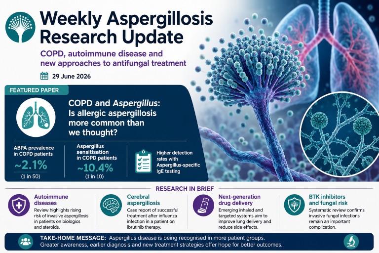
COPD, autoimmune disease and new approaches to antifungal treatment
Published: 29 June 2026
Every week we review the latest research on aspergillosis and related fungal diseases, selecting the studies most likely to influence patient care and clinical practice. This week's research focuses on an increasingly important theme: recognising Aspergillus disease in patient groups where it has traditionally been overlooked.
The highlight is a large systematic review suggesting that allergic Aspergillus disease may be more common in people living with chronic obstructive pulmonary disease (COPD) than previously appreciated.
Featured Paper
COPD and Aspergillus: Is allergic aspergillosis more common than we thought?
For decades, allergic bronchopulmonary aspergillosis (ABPA) has been regarded primarily as a complication of asthma and cystic fibrosis. However, respiratory specialists have increasingly reported Aspergillus-related disease in patients with COPD.
A new systematic review and meta-analysis has now brought together the available evidence.
Paper: Ajayababu A, Antony A, Goyal B, Ray A. Prevalence of allergic bronchopulmonary aspergillosis/Aspergillus sensitization in chronic obstructive pulmonary disease: A systematic review and meta-analysis. Respiratory Investigation. 2026. PubMed PMID: 42361722
What did the researchers do?
The authors searched four major medical databases for studies reporting either:
- Allergic bronchopulmonary aspergillosis (ABPA)
- Aspergillus sensitisation (AS)
Among patients with COPD, the review identified 23 suitable studies, including:
- 1,529 patients for analysis of ABPA
- 3,505 patients for analysis of Aspergillus sensitisation
Pooling data from many studies provides a more reliable estimate than individual reports alone.
What did they find?
The results suggest Aspergillus-related disease is not rare in COPD.
The pooled prevalence was:
- ABPA: 2.1%
- Aspergillus sensitisation: 10.4%
Put another way:
- around 1 in 50 people with COPD may have ABPA
- approximately 1 in 10 have evidence of sensitisation to Aspergillus
Studies using Aspergillus-specific IgE as part of their diagnostic strategy detected more ABPA than studies using skin testing or total IgE alone. This suggests that the choice of diagnostic tests may influence how many patients are identified.
Why is this important?
Many symptoms of COPD overlap with Aspergillus-related disease, including:
- persistent cough
- breathlessness
- increased sputum production
- recurrent exacerbations
As a result, some patients may continue to receive repeated courses of antibiotics or steroids while an underlying Aspergillus-related condition remains unrecognised.
This study does not suggest that everyone with COPD should undergo routine fungal testing. However, it supports considering Aspergillus investigations in selected patients with:
- recurrent exacerbations despite optimal therapy
- unexplained eosinophilia or raised IgE
- bronchiectasis
- persistent mucus plugging
- radiological abnormalities that do not fit the expected pattern
Strengths and limitations
This is currently one of the largest reviews examining COPD and Aspergillus disease.
Its strengths include:
- systematic literature search
- formal meta-analysis
- inclusion of more than 5,000 patients overall
However, the included studies differed considerably in patient populations, diagnostic methods and disease severity. This statistical heterogeneity means the true prevalence almost certainly varies between different clinical settings.
The review also demonstrates an association rather than proving that COPD itself causes ABPA.
What does this mean for patients?
The main message is one of greater awareness.
For patients with COPD whose symptoms remain difficult to explain despite appropriate treatment, clinicians may increasingly consider whether Aspergillus sensitisation or ABPA could be contributing to ongoing respiratory problems.
Earlier recognition has the potential to improve diagnosis and ensure that patients receive the most appropriate investigations and treatment.
Research in Brief
Invasive aspergillosis in autoimmune inflammatory rheumatic diseases
Paper: Liang P, Zhang X, Cai S, Hu Z, Dong L. Invasive aspergillosis in autoimmune inflammatory rheumatic diseases: epidemiology, risk factors, diagnosis, management and challenges. Annals of Medicine. 2026. PubMed PMID: 42343869
A comprehensive new review highlights the growing importance of invasive aspergillosis in people with autoimmune inflammatory rheumatic diseases such as rheumatoid arthritis and systemic vasculitis.
The increasing use of corticosteroids, biologic therapies and other immunosuppressive medications has expanded the population at risk. The authors note that invasive aspergillosis remains uncommon but carries a high mortality when diagnosis is delayed.
A particular challenge is that symptoms and imaging findings can resemble a flare of the underlying autoimmune disease, making diagnosis difficult. The review emphasises combining clinical assessment with microbiological tests, including galactomannan, PCR and bronchoalveolar lavage where appropriate, and careful interpretation of imaging findings.
Why it matters: Clinicians should maintain a high index of suspicion for invasive fungal disease in immunosuppressed patients who fail to respond as expected to conventional treatment.
Cerebral aspergillosis following influenza and ibrutinib therapy
Paper: Haraguchi M, Kimura M, Uruga H, Takahashi Y, Takaya H, Arisawa K, et al. Cerebral aspergillosis caused by Aspergillus flavus following seasonal influenza infection in a patient receiving ibrutinib for Waldenström's macroglobulinemia. Journal of Infection and Chemotherapy. 2026. PubMed PMID: 42331321
A Japanese case report describes successful treatment of cerebral aspergillosis caused by Aspergillus flavus in a patient receiving the Bruton tyrosine kinase inhibitor ibrutinib after seasonal influenza infection.
The patient developed both pulmonary and cerebral aspergillosis, underwent neurosurgical debridement and was successfully treated with isavuconazole.
Although this represents a single case, it reinforces growing evidence that BTK inhibitors and severe viral infections are emerging risk factors for invasive aspergillosis.
Why it matters: Patients receiving targeted therapies who develop persistent respiratory or neurological symptoms following influenza warrant careful assessment for opportunistic fungal infection.
Looking ahead: New ways to deliver antifungal drugs
Paper: Martins YA, Anselmo-Lima WT, Tamashiro E, Ho E, Valera FCP. Next-generation drug delivery systems for aspergillosis: Overcoming barriers in antifungal therapy. Biomedicine & Pharmacotherapy. 2026. PubMed PMID: 42361622
Another review this week explores next-generation drug delivery systems designed to improve treatment of aspergillosis.
Researchers are developing inhaled formulations, nanoparticles and targeted drug-delivery technologies that aim to increase antifungal concentrations directly within the lungs while reducing systemic side effects.
Most of these approaches remain experimental, but they offer possibilities for the future management of chronic pulmonary aspergillosis and other forms of pulmonary fungal disease.
What this week's research tells us
Although these papers address different aspects of aspergillosis, they all point in the same direction.
Increasingly, Aspergillus disease is being recognised in patient groups previously considered to be at relatively low risk, including people with COPD and those receiving modern immunosuppressive therapies.
At the same time, advances in diagnostics and drug delivery are creating opportunities for earlier diagnosis and more targeted treatment.
For patients, the message is reassuring but important: persistent or unexplained respiratory symptoms deserve careful evaluation, particularly when standard treatments are not achieving the expected improvement.
As awareness continues to grow, more patients may receive the correct diagnosis earlier in the course of their illness.
References
- Ajayababu A, Antony A, Goyal B, Ray A. Prevalence of allergic bronchopulmonary aspergillosis/Aspergillus sensitization in chronic obstructive pulmonary disease: A systematic review and meta-analysis. Respiratory Investigation. 2026. doi:10.1016/j.resinv.2026.101469. PubMed
- Liang P, Zhang X, Cai S, Hu Z, Dong L. Invasive aspergillosis in autoimmune inflammatory rheumatic diseases: epidemiology, risk factors, diagnosis, management and challenges. Annals of Medicine. 2026. doi:10.1080/07853890.2026.2685285. PubMed
- Haraguchi M, Kimura M, Uruga H, Takahashi Y, Takaya H, Arisawa K, et al. Cerebral aspergillosis caused by Aspergillus flavus following seasonal influenza infection in a patient receiving ibrutinib for Waldenström's macroglobulinemia. Journal of Infection and Chemotherapy. 2026. doi:10.1016/j.jiac.2026.103020. PubMed
- Martins YA, Anselmo-Lima WT, Tamashiro E, Ho E, Valera FCP. Next-generation drug delivery systems for aspergillosis: Overcoming barriers in antifungal therapy. Biomedicine & Pharmacotherapy. 2026. doi:10.1016/j.biopha.2026.119691. PubMed
```

Professional Aspergillosis Update: May 2026
Audience: respiratory physicians, infectious diseases physicians, clinical microbiologists, haematologists, pharmacists, specialist nurses, laboratory scientists and researchers with an interest in aspergillosis.
Contents
Key messages
- Isavuconazole therapeutic drug monitoring may have a selective role. Although isavuconazole is usually more predictable than voriconazole, real-world pharmacokinetic variability remains clinically relevant in some patients.
- Posaconazole prophylaxis should not automatically be avoided with midostaurin. The interaction is real, but clinical consequences may often be manageable with careful monitoring.
- Surrogate azole susceptibility testing has limits. Voriconazole gradient diffusion testing may help screen for broader azole resistance, but it should not replace direct susceptibility testing where treatment decisions depend on the result.
- Invasive fungal sinusitis remains a high-mortality emergency in haematological malignancy. Early tissue diagnosis, ENT involvement and multidisciplinary management remain central.
- Non-fumigatus Aspergillus species are becoming more important research targets. New CRISPR-Cas9 tools for Aspergillus calidoustus may support future work on virulence and antifungal resistance.
Top papers this month
1. Isavuconazole pharmacokinetics and pharmacodynamics in real-world practice
Guidi M, Couchepin J, Reinhold I, Kronig I, Neofytos D, Schreiber PW, André P, Buclin T, Lamoth F.
Characterization of isavuconazole pharmacokinetics and pharmacodynamics in a real-life cohort.
JAC Antimicrobial Resistance. 2026;8(3):dlag071.
PMID: 42088097
Why this paper was selected
Isavuconazole is increasingly used for invasive aspergillosis because of its favourable safety profile and generally more predictable pharmacokinetics compared with voriconazole. This study provides important real-world evidence that clinically relevant interpatient variability still occurs and that therapeutic drug monitoring may have a role in selected patients.
Key findings
- Isavuconazole showed relatively predictable pharmacokinetics overall.
- Clinically relevant variability in drug exposure was still observed between patients.
- Therapeutic drug monitoring identified patients with atypically low or high exposure.
- Exposure relative to fungal minimum inhibitory concentration may be more informative than plasma concentration alone.
- No strong concentration-dependent toxicity signal was observed within the exposure range studied.
Clinical significance
This paper challenges the assumption that isavuconazole therapeutic drug monitoring is rarely useful. While the findings do not justify universal routine monitoring, they support selective monitoring in complex patients, particularly where there is treatment failure, suspected malabsorption, significant drug interactions, unusual body composition, long-term therapy, or infection with isolates showing elevated minimum inhibitory concentrations.
Implications for practice
Classification: Important but not yet practice changing.
The study supports a more individualised approach to isavuconazole use. It also reinforces the direction of travel in antifungal stewardship: interpreting drug exposure alongside fungal susceptibility rather than considering plasma concentrations in isolation.
Evidence assessment
Evidence quality: Moderate. The real-world dataset and pharmacokinetic-pharmacodynamic modelling strengthen the evidence base, but the observational design limits causal inference and definitive exposure targets were not established.
Editorial assessment
This is one of the most clinically relevant antifungal pharmacology papers in this update. It does not establish mandatory isavuconazole monitoring, but it provides a strong argument for selective therapeutic drug monitoring in high-risk or complex aspergillosis patients.
2. Managing posaconazole and midostaurin interactions in FLT3-mutated AML
Joisten CS, Mellinghoff SC, Seidel D, Müller C, Müller-Ohrem C, Kreuzer K-A, Frenzel LP, Simon F, Hallek M, Koehler P, Cornely OA, Stemler J.
Clinical impact of potential drug-drug interactions between midostaurin and posaconazole in FLT3-mutated AML.
Antimicrobial Agents and Chemotherapy. 2026;70(6):e01951-25.
PMID: 42118097
Why this paper was selected
Posaconazole prophylaxis is central to prevention of invasive aspergillosis in patients undergoing intensive acute myeloid leukaemia treatment. Midostaurin is metabolised through CYP3A4, and posaconazole is a potent CYP3A4 inhibitor. This study addresses a common real-world dilemma: whether this interaction should alter antifungal prophylaxis practice.
Key findings
- The pharmacokinetic interaction between posaconazole and midostaurin was confirmed.
- Clinical toxicity appeared less severe than theoretical concerns might suggest.
- Many patients were able to receive both agents without major treatment-limiting toxicity.
- Individual variability in exposure and tolerability remained important.
- The findings support continued attention to monitoring rather than automatic avoidance of posaconazole.
Clinical significance
This paper is important because it addresses an immediate bedside decision. Avoiding posaconazole because of interaction concerns may leave high-risk acute myeloid leukaemia patients vulnerable to invasive aspergillosis. The study suggests that the interaction is clinically manageable in many patients when appropriate monitoring and multidisciplinary oversight are in place.
Implications for practice
Classification: Important but not yet practice changing.
The paper supports continued use of posaconazole prophylaxis where clinically indicated, with careful monitoring for toxicity and close collaboration between haematology, infectious diseases, microbiology and pharmacy teams.
Evidence assessment
Evidence quality: Moderate. The study is clinically relevant and real-world, but observational. It does not establish definitive dose-adjustment protocols or replace existing guideline recommendations.
Editorial assessment
The key message is that proven antifungal prophylaxis should not be abandoned solely because of theoretical interaction concerns. The interaction is real, but careful monitoring is generally preferable to withholding protection against invasive aspergillosis in a very high-risk group.
3. Can voriconazole susceptibility predict isavuconazole or posaconazole susceptibility?
Vahedi-Shahandashti R, Nickel A-S, Eisele D, Lass-Flörl C; ISHAM Working Group Member of Intrinsic Antifungal Resistance.
Can voriconazole gradient diffusion testing results be extrapolated to isavuconazole and posaconazole in Aspergillus spp.? Comparative analysis with CLSI broth microdilution and cyp51A gene sequencing.
Antimicrobial Agents and Chemotherapy. 2026;70(6):e01813-25.
PMID: 42138696
Why this paper was selected
Azole resistance in Aspergillus species is a growing problem, but not all laboratories can perform comprehensive susceptibility testing for every triazole. This paper asks whether voriconazole gradient diffusion testing can be used as a practical surrogate marker for broader azole susceptibility.
Key findings
- Voriconazole susceptibility often correlated with broader azole susceptibility patterns.
- Elevated voriconazole minimum inhibitory concentrations frequently corresponded with reduced isavuconazole susceptibility.
- Prediction of posaconazole susceptibility was less reliable.
- Discordant susceptibility profiles occurred, particularly among resistant isolates.
- cyp51A sequencing helped explain many resistance patterns but did not account for all phenotypes.
Clinical significance
The study supports voriconazole gradient diffusion testing as a useful first-line screening approach, especially where full reference testing is not immediately available. However, it also highlights a critical limitation: susceptibility to one triazole cannot be assumed to guarantee susceptibility to another.
Implications for practice
Classification: Important but not yet practice changing.
Voriconazole gradient diffusion testing may help identify isolates that require further investigation, but it should not replace direct isavuconazole or posaconazole susceptibility testing where treatment decisions depend on accurate results.
Evidence assessment
Evidence quality: Moderate to high for a laboratory diagnostic study. The use of CLSI broth microdilution and cyp51A sequencing strengthens the analysis, but clinical outcome data were not assessed.
Editorial assessment
This is a practical paper for clinical mycology laboratories. The main message is that surrogate azole testing can support screening and stewardship, but definitive treatment decisions should still be based on agent-specific susceptibility testing and molecular resistance analysis where available.
4. Invasive fungal sinusitis in haematological malignancy
Athni TS, Strauch CB, Kovac V, Arbona-Haddad E, Villa IP, Gupta S, Aleissa MM, Liakos AD, Tong A, Vedula RS, Maxfield AZ, Bergmark RW, Sherman AC.
Invasive fungal sinusitis in patients with hematological malignancies: a 20-year study from a tertiary academic US hospital system.
Open Forum Infectious Diseases. 2026;13(6):ofag304.
PMID: 42238379
Why this paper was selected
Invasive fungal sinusitis is a severe but less commonly discussed manifestation of invasive mould disease. In haematological malignancy, delayed recognition can lead to orbital, intracranial and fatal complications. This 20-year cohort provides useful long-term clinical insight.
Key findings
- Aspergillus species and Mucorales were the dominant pathogens.
- Mortality remained substantial despite modern antifungal therapy and supportive care.
- Early imaging, endoscopic assessment, tissue biopsy and histopathology remained central to diagnosis.
- Successful management frequently required combined medical and surgical approaches.
- Multidisciplinary care involving haematology, infectious diseases, ENT, microbiology and radiology was essential.
Clinical significance
This study reinforces that invasive aspergillosis is not solely a pulmonary disease. Sinonasal invasive fungal disease remains an emergency in profoundly immunocompromised patients. Distinguishing aspergillosis from mucormycosis is particularly important because antifungal treatment choices differ substantially.
Implications for practice
Classification: Important but not practice changing.
The paper reinforces existing best practice: early suspicion, urgent ENT involvement, tissue diagnosis, prompt antifungal therapy and multidisciplinary management.
Evidence assessment
Evidence quality: Moderate. The long observation period and detailed clinical experience are strengths, but the retrospective single-system design limits causal conclusions.
Editorial assessment
This paper is a useful reminder that early recognition remains one of the strongest determinants of outcome in invasive fungal disease. Persistent or atypical sinus symptoms in high-risk haematology patients should prompt urgent assessment rather than routine treatment as uncomplicated bacterial sinusitis.
Important development
5. Invasive mould infections in transplant recipients
Sudhaharan S, Pamidimukkala U, Bojja S, Raju DSB, Kk R, Gopal PSS.
Invasive mold infections among transplant recipients: a single-center observational study.
Journal de Mycologie Médicale / Journal of Medical Mycology. 2026;36(2):101629.
DOI: 10.1016/j.mycmed.2026.101629
Why this paper was selected
Transplant recipients remain a key high-risk population for invasive aspergillosis and other invasive mould infections. This observational study provides contemporary real-world data on presentation, diagnosis, microbiology, treatment and outcomes in a transplant centre.
Key findings
- Aspergillus species remained the predominant mould pathogen.
- Pulmonary disease was the most common presentation.
- Diagnosis required multimodal assessment combining clinical, radiological and mycological data.
- Invasive mould infections remained associated with substantial morbidity and mortality.
- Earlier diagnosis was associated with more favourable outcomes.
Clinical significance
The study confirms rather than changes current understanding. Its main value is as a contemporary reminder that invasive aspergillosis remains a major threat in transplantation despite advances in prophylaxis, diagnostics and antifungal treatment.
Implications for practice
Classification: Important but not practice changing.
The findings support ongoing vigilance, rapid investigation pathways, early multidisciplinary input and antifungal stewardship in transplant programmes.
Evidence assessment
Evidence quality: Moderate. Real-world applicability is useful, but the single-centre observational design and modest sample size limit generalisability.
Editorial assessment
This paper does not introduce a new management strategy, but it reinforces an enduring message: invasive aspergillosis outcomes in transplant recipients remain strongly dependent on early recognition and timely treatment.
Research horizon
6. CRISPR-Cas9 gene editing in Aspergillus calidoustus
Hollomon JM, Dahlstrom KM.
CRISPR-Cas9-mediated targeted gene deletion in Aspergillus calidoustus, a non-model environmental mold.
Microbiology Spectrum. 2026;14(6):e03899-25.
PMID: 42112836
Why this paper was selected
Most molecular understanding of pathogenic Aspergillus species comes from Aspergillus fumigatus. This study establishes a CRISPR-Cas9 gene-editing system for Aspergillus calidoustus, an emerging opportunistic mould with clinical relevance and reduced susceptibility to some antifungals.
Key findings
- The authors successfully developed a CRISPR-Cas9 platform for targeted gene deletion in A. calidoustus.
- The system provides a method for functional genetic studies in a previously less tractable species.
- The platform may support future research into virulence, environmental adaptation, antifungal resistance and novel drug targets.
Clinical significance
There is no immediate clinical application. However, the study is important as enabling science. As non-fumigatus Aspergillus species are increasingly recognised in clinical practice, tools that allow their biology to be studied directly may become increasingly valuable.
Implications for practice
Classification: Early-stage research requiring further validation.
This paper does not alter clinical management, diagnostics or guidelines. Its value lies in supporting future translational research.
Editorial assessment
This is a foundational research paper. It will not change patient care today, but it may help build the scientific infrastructure needed to understand emerging mould pathogens and their resistance mechanisms over the next decade.
Clinical pearl
7. Primary traumatic cutaneous aspergillosis caused by Aspergillus terreus
Ing SK, Lee YH, Tan YY, Aziz MBA, Chang AKW.
Primary traumatic cutaneous aspergillosis of the hand caused by Aspergillus terreus following a mould-contaminated penetrating injury.
Medical Mycology Case Reports. 2026;52:100798.
PMID: 42237979
Why this case was noted
This case report describes primary traumatic cutaneous aspergillosis of the hand caused by Aspergillus terreus following a mould-contaminated penetrating injury.
Clinical take-home points
- Aspergillosis is not always acquired through inhalation.
- Direct traumatic inoculation can cause localised Aspergillus infection.
- Persistent or atypical wounds following mould-contaminated trauma should prompt consideration of fungal infection.
- Tissue sampling is essential for diagnosis.
- Species-level identification matters because Aspergillus terreus is intrinsically resistant to amphotericin B.
Editorial assessment
This is not a practice-changing paper, but it is a useful educational case. It broadens clinical awareness beyond pulmonary aspergillosis and highlights the importance of early tissue diagnosis when wounds behave unexpectedly after contaminated trauma.
Overall editorial summary
The May 2026 literature contains several papers that are useful for clinicians and laboratory professionals working in aspergillosis and invasive mould disease. The strongest clinical themes are antifungal stewardship, drug exposure, azole resistance, and the continued importance of early diagnosis in high-risk populations.
The isavuconazole pharmacokinetic-pharmacodynamic study and the midostaurin-posaconazole interaction paper are particularly relevant because they address practical treatment decisions. The azole susceptibility study is highly relevant to clinical mycology laboratories and reinforces the need for careful interpretation of surrogate resistance testing. The invasive fungal sinusitis and transplant studies reinforce a familiar but important message: outcomes remain closely linked to early recognition, tissue diagnosis where appropriate, and multidisciplinary management.
Finally, the CRISPR-Cas9 paper and traumatic cutaneous aspergillosis case illustrate the breadth of modern aspergillosis research, from molecular tools for emerging moulds to unusual clinical presentations outside the respiratory tract.
References
- Guidi M, Couchepin J, Reinhold I, Kronig I, Neofytos D, Schreiber PW, André P, Buclin T, Lamoth F. Characterization of isavuconazole pharmacokinetics and pharmacodynamics in a real-life cohort. JAC Antimicrobial Resistance. 2026;8(3):dlag071. PMID: 42088097
- Joisten CS, Mellinghoff SC, Seidel D, Müller C, Müller-Ohrem C, Kreuzer K-A, Frenzel LP, Simon F, Hallek M, Koehler P, Cornely OA, Stemler J. Clinical impact of potential drug-drug interactions between midostaurin and posaconazole in FLT3-mutated AML. Antimicrobial Agents and Chemotherapy. 2026;70(6):e01951-25. PMID: 42118097
- Vahedi-Shahandashti R, Nickel A-S, Eisele D, Lass-Flörl C; ISHAM Working Group Member of Intrinsic Antifungal Resistance. Can voriconazole gradient diffusion testing results be extrapolated to isavuconazole and posaconazole in Aspergillus spp.? Comparative analysis with CLSI broth microdilution and cyp51A gene sequencing. Antimicrobial Agents and Chemotherapy. 2026;70(6):e01813-25. PMID: 42138696
- Athni TS, Strauch CB, Kovac V, Arbona-Haddad E, Villa IP, Gupta S, Aleissa MM, Liakos AD, Tong A, Vedula RS, Maxfield AZ, Bergmark RW, Sherman AC. Invasive fungal sinusitis in patients with hematological malignancies: a 20-year study from a tertiary academic US hospital system. Open Forum Infectious Diseases. 2026;13(6):ofag304. PMID: 42238379
- Sudhaharan S, Pamidimukkala U, Bojja S, Raju DSB, Kk R, Gopal PSS. Invasive mold infections among transplant recipients: a single-center observational study. Journal de Mycologie Médicale / Journal of Medical Mycology. 2026;36(2):101629. DOI: 10.1016/j.mycmed.2026.101629
- Hollomon JM, Dahlstrom KM. CRISPR-Cas9-mediated targeted gene deletion in Aspergillus calidoustus, a non-model environmental mold. Microbiology Spectrum. 2026;14(6):e03899-25. PMID: 42112836
- Ing SK, Lee YH, Tan YY, Aziz MBA, Chang AKW. Primary traumatic cutaneous aspergillosis of the hand caused by Aspergillus terreus following a mould-contaminated penetrating injury. Medical Mycology Case Reports. 2026;52:100798. PMID: 42237979
Article information
Prepared for: aspergillosis.org professionals section
Intended audience: healthcare professionals and researchers
Article type: monthly professional literature update
Coverage period: May 2026
Last reviewed: June 2026
Specialist Evidence Briefing: Aspergillus Serology for CPA Diagnosis: Why Assay Choice, Cut-Offs and Confirmatory Testing Matter
Key points
- CPA diagnosis remains challenging because symptoms and radiology overlap with tuberculosis, non-tuberculous mycobacterial disease, bronchiectasis, chronic obstructive pulmonary disease, malignancy and other chronic lung disorders.
- Aspergillus immunoglobulin G (IgG) testing is a cornerstone of CPA diagnosis, but performance varies between assays.
- Cut-off selection affects sensitivity and specificity, with important consequences for both missed CPA and overdiagnosis.
- Confirmatory or complementary testing, such as Western blot in selected situations, may help clarify difficult or borderline cases.
- The paper is directly relevant to specialist respiratory, infectious diseases and mycology services because diagnostic reliability affects referral pathways, antifungal prescribing and case definition.
The paper
Bigot J, Gibert C, Millet N, et al. Aspergillus serology for chronic pulmonary aspergillosis diagnosis: optimization of an enzyme-linked immunosorbent assay kit and assessment of a Western blot kit performance. Journal of Clinical Microbiology. Published 28 May 2026. doi:10.1128/jcm.00182-26.
Why this paper matters
CPA is a progressive and potentially fatal chronic lung infection that usually occurs in patients with underlying structural lung damage. Diagnosis depends on combining clinical symptoms, characteristic radiology and evidence of Aspergillus infection or immune response.
In practice, Aspergillus serology is often the decisive test. A positive Aspergillus IgG result can support the diagnosis when imaging and symptoms are compatible. A negative result can reduce diagnostic probability. But serology is not absolute. Different assays use different antigen preparations, platforms and reporting units. This means that results from one laboratory may not be directly comparable with results from another.
This paper is therefore important because it addresses a real-world diagnostic problem: how to optimise Aspergillus serological testing so that it performs reliably in the diagnosis of CPA.
Clinical background: why CPA serology is difficult
CPA often develops in patients with structurally abnormal lungs. Common underlying conditions include previous pulmonary tuberculosis, non-tuberculous mycobacterial infection, chronic obstructive pulmonary disease, bronchiectasis, sarcoidosis, prior lung surgery and other cavitary lung diseases.
These same conditions can also cause chronic symptoms and abnormal imaging without CPA. This creates a diagnostic grey zone. Patients may have cavities, pleural thickening, fibrosis, bronchiectasis, chronic cough, haemoptysis or weight loss for reasons other than Aspergillus disease.
Aspergillus IgG testing is therefore valuable because it provides evidence of an immune response to Aspergillus. However, it must be interpreted in context. A positive result does not, by itself, prove active CPA. A negative result does not always fully exclude CPA, particularly if clinical suspicion remains high.
What the study evaluates
The paper evaluates two related aspects of CPA serology:
- Optimisation of an enzyme-linked immunosorbent assay (ELISA) kit for Aspergillus serology in CPA diagnosis.
- Assessment of Western blot kit performance, potentially as a complementary or confirmatory approach.
This is clinically relevant because ELISA-based Aspergillus IgG testing is widely used in diagnostic pathways, while Western blot may offer additional qualitative information in selected cases. The practical question is whether assay optimisation and confirmatory testing improve diagnostic performance enough to change specialist practice.
Why assay cut-offs matter
The choice of diagnostic cut-off is not a technical detail; it changes clinical classification.
| Cut-off strategy | Likely effect | Clinical risk |
|---|---|---|
| Lower cut-off | Higher sensitivity | More false positives and possible overdiagnosis |
| Higher cut-off | Higher specificity | More false negatives and missed CPA |
| Population-specific cut-off | Potentially better clinical performance | May reduce comparability between centres |
| Single universal cut-off | Simpler implementation | May perform poorly across different populations |
In a specialist CPA service, false negatives may delay antifungal treatment and referral. False positives may lead to unnecessary anxiety, additional imaging, prolonged antifungal therapy and avoidable toxicity. The correct balance depends on the clinical setting, disease prevalence and consequences of diagnostic error.
Where Western blot may fit
Western blot testing may be useful where ELISA results are borderline, discordant with imaging, or difficult to interpret in patients with complex lung disease. It may also be useful as a complementary method in diagnostic algorithms, depending on availability and validated performance.
However, Western blot should not be seen as a simple replacement for ELISA. Its value depends on whether it adds diagnostic clarity beyond standard serology, whether it is reproducible between laboratories, and whether clinicians know how to act on the result.
Clinical interpretation: serology is supportive, not standalone
For specialists, the key principle remains:
Aspergillus serology supports the diagnosis of CPA but does not diagnose CPA in isolation.
A robust CPA diagnosis requires integration of:
- compatible symptoms, usually chronic cough, weight loss, fatigue, breathlessness or haemoptysis;
- compatible imaging, particularly cavities, pleural thickening, pericavitary infiltrates, fungal ball, nodules or progressive fibrosis;
- mycological or immunological evidence, especially Aspergillus IgG;
- exclusion or recognition of alternative and co-existing diagnoses, including active tuberculosis, non-tuberculous mycobacterial disease, malignancy and bacterial bronchiectasis.
Implications for specialist services
This paper supports the need for carefully governed CPA serology pathways. Specialist centres and diagnostic laboratories should consider:
- which Aspergillus IgG assay is used locally;
- what cut-off is applied and how it was validated;
- whether borderline zones should be reported rather than simple positive/negative categories;
- how results are interpreted in high-risk structural lung disease populations;
- whether confirmatory testing is available for selected cases;
- how serology is integrated with CT findings and microbiology;
- whether local reports include interpretive comments to reduce misuse.
Suggested diagnostic reporting approach
Rather than reporting Aspergillus serology as a binary answer, laboratories and specialist services may benefit from a more interpretive model:
| Result category | Possible interpretation | Suggested action |
|---|---|---|
| Clearly negative | CPA less likely, but not impossible if clinical suspicion is high | Review imaging and alternative diagnoses; repeat if disease evolves |
| Borderline or low positive | Uncertain significance | Correlate with CT, symptoms, cultures and prior results; consider repeat or confirmatory testing |
| Clearly positive | Supports CPA in the right clinical/radiological context | Assess full CPA criteria and consider specialist referral or treatment discussion |
| Discordant result | Serology conflicts with clinical picture | Reassess diagnosis, assay limitations and possibility of co-existing disease |
Relevance to post-tuberculosis lung disease
This paper is particularly relevant when considered alongside recent evidence that CPA may be detected during or soon after pulmonary tuberculosis treatment. In post-tuberculosis lung disease, cavities and chronic radiological abnormalities are common, but not all symptoms are due to active TB or permanent scarring.
Reliable Aspergillus serology is therefore essential. If the assay cut-off is poorly calibrated, clinicians may either miss CPA in symptomatic patients with cavities or overdiagnose CPA in patients with structural lung damage but no active Aspergillus disease.
Relevance to bronchiectasis and non-tuberculous mycobacterial disease
Patients with bronchiectasis or non-tuberculous mycobacterial lung disease may also have chronic symptoms, mucus production, recurrent infection, radiological change and occasional Aspergillus isolation. Aspergillus serology can be useful in identifying CPA, but positive results must be interpreted carefully because these patients often have complex chronic airway disease.
For this group, assay performance and cut-off choice may substantially affect diagnostic confidence.
Implications for antifungal stewardship
Improved CPA serology has direct stewardship implications. Antifungal treatment for CPA is often prolonged and requires monitoring for toxicity, drug interactions and therapeutic drug levels. Overdiagnosis can expose patients to unnecessary azoles. Underdiagnosis can allow progressive cavitary disease, haemoptysis and loss of lung function.
More reliable serological testing can therefore support both earlier treatment in true CPA and avoidance of unnecessary therapy in patients who do not meet diagnostic criteria.
Evidence strength
| Question | Evidence strength | Comment |
|---|---|---|
| Is Aspergillus IgG central to CPA diagnosis? | Strong | Recognised in major CPA diagnostic guidance |
| Do different assays and cut-offs affect performance? | Strong | Well-recognised practical issue in CPA serology |
| Can assay optimisation improve diagnostic classification? | Likely | This paper directly addresses optimisation, but local validation remains important |
| Should Western blot replace ELISA? | Not established | More likely to be complementary or confirmatory in selected cases |
| Should serology alone determine CPA treatment? | No | Must be interpreted with symptoms, imaging and microbiology |
Practical take-home messages for specialists
- Know which Aspergillus IgG assay your laboratory uses.
- Know the cut-off and whether it has been validated for CPA.
- Be cautious with borderline results.
- Do not diagnose CPA on serology alone.
- Do not dismiss CPA solely because one serological test is negative if the clinical and CT picture is highly suggestive.
- Consider repeat or complementary testing when results are discordant.
- Integrate serology with CT, symptoms, sputum fungal culture/PCR and assessment for tuberculosis or non-tuberculous mycobacteria.
Conclusion
This paper is a timely reminder that CPA diagnosis depends not only on whether Aspergillus serology is performed, but on how well the assay is optimised, how cut-offs are selected and how results are interpreted. For specialist services, improved serology can strengthen diagnostic confidence, support earlier recognition of CPA and reduce unnecessary antifungal treatment.
The broader implication is clear: CPA diagnostic pathways need standardised, validated and clinically interpreted Aspergillus serology, not isolated positive or negative blood test results.
References
- Bigot J, Gibert C, Millet N, et al. Aspergillus serology for chronic pulmonary aspergillosis diagnosis: optimization of an enzyme-linked immunosorbent assay kit and assessment of a Western blot kit performance.
Journal of Clinical Microbiology. Published 28 May 2026.
doi:10.1128/jcm.00182-26.
PubMed - Denning DW, Cadranel J, Beigelman-Aubry C, et al. Chronic pulmonary aspergillosis: rationale and clinical guidelines for diagnosis and management.
European Respiratory Journal. 2016;47(1):45-68.
doi:10.1183/13993003.00583-2015.
PubMed - Patterson TF, Thompson GR, Denning DW, et al. Practice guidelines for the diagnosis and management of aspergillosis: 2016 update by the Infectious Diseases Society of America.
Clinical Infectious Diseases. 2016;63(4):e1-e60.
doi:10.1093/cid/ciw326.
PubMed
Commercial Aspergillus fumigatus real-time PCR for invasive pulmonary aspergillosis: specialist evidence briefing
Why this paper matters
Invasive pulmonary aspergillosis remains difficult to diagnose early, particularly in patients with haematological malignancy, haematopoietic stem cell transplantation, intensive chemotherapy, corticosteroid exposure or profound immunosuppression. Culture is insensitive, histology is often unavailable, and radiology is not specific. Galactomannan has become a central biomarker, but performance varies by specimen type, antifungal exposure, host group and disease stage.
Aspergillus PCR has long been promising, but implementation has been limited by assay heterogeneity, extraction differences, target selection, contamination concerns, and variable interpretation across centres. A clinically evaluated commercial real-time PCR assay is therefore important because commercialisation may improve standardisation, reproducibility and adoption outside specialist mycology laboratories.
Clinical context
The diagnosis of IPA is usually probabilistic. Clinicians combine:
- host factors, such as neutropenia, haematological malignancy, transplantation or corticosteroid exposure;
- compatible imaging, especially nodules, halo sign, cavitation or infarct-like lesions on computed tomography;
- mycological evidence, including galactomannan, culture, microscopy, PCR or lateral-flow assays;
- clinical course and response to antifungal therapy.
In this setting, a commercial Aspergillus fumigatus real-time PCR assay could support earlier diagnosis, improve confidence in probable IPA classification, and potentially reduce unnecessary empirical antifungal treatment when used as part of a diagnostic algorithm.
Main finding
The paper evaluates a commercial Aspergillus fumigatus real-time PCR assay for the diagnosis of invasive pulmonary aspergillosis in patients with haematological malignancies. The clinical relevance lies in validating a defined assay in a high-risk population where early diagnosis directly affects antifungal timing and survival.
The specialist significance is not simply that PCR “works”. The key question is whether a commercial PCR assay offers sufficiently reliable analytical and clinical performance to be used alongside galactomannan and imaging in routine pathways.
What is genuinely new?
The novelty is likely to sit in one or more of the following areas:
- evaluation of a specific commercial assay rather than an in-house laboratory-developed test;
- clinical validation in a defined haematology population;
- comparison against established diagnostic categories such as proven/probable IPA;
- assessment of analytical performance, including limit of detection, reproducibility or specificity;
- potential contribution to standardisation of Aspergillus PCR implementation.
This is not a new concept in principle: Aspergillus PCR has been studied for many years. What is more clinically useful is the transition from heterogeneous in-house assays towards assays that can be validated, quality-assured and compared across centres.
Relationship to existing evidence
Recent reviews of molecular fungal diagnostics emphasise that PCR can improve the diagnosis of invasive fungal disease but remains limited by standardisation, assay availability and interpretation. A 2025 review in Diagnostics noted that PCR is sensitive and specific for invasive fungal disease, but that implementation remains constrained by limited standardisation, few commercial options, and lack of clear guidance for interpreting results.
That surrounding evidence makes this type of paper important. Commercial PCR assays do not automatically solve diagnostic uncertainty, but they can reduce one major barrier: between-laboratory variability.
Strengths to look for in the paper
- Clearly defined patient population, especially haematological malignancy or transplant subgroups.
- Use of accepted case definitions for proven, probable and possible IPA.
- Separate analytical and clinical performance assessment.
- Comparison with galactomannan, culture and radiology.
- Evaluation by specimen type, especially bronchoalveolar lavage fluid versus blood.
- Consideration of antifungal exposure before sampling.
- Reporting of sensitivity, specificity, positive predictive value and negative predictive value.
Limitations and cautions
PCR performance depends strongly on specimen type. Bronchoalveolar lavage fluid generally provides a higher organism burden than blood in IPA, but it is more invasive and may not be feasible in unstable or thrombocytopenic patients. Blood PCR is less invasive but may be less sensitive, especially in localised airway-invasive disease.
False positives may arise from colonisation, contamination or detection of non-invasive airway presence. False negatives may occur with low fungal burden, prior antifungal therapy, sampling timing, extraction inefficiency or inhibitors. A positive PCR result should therefore not be interpreted in isolation.
A further issue is species coverage. A narrowly targeted Aspergillus fumigatus assay may perform well for A. fumigatus but could miss non-fumigatus Aspergillus species or cryptic species with different susceptibility patterns. In haematology patients, that may matter for epidemiology and antifungal resistance surveillance.
Clinical implications
For specialist services, the likely implication is that commercial Aspergillus PCR should be considered as part of a multi-modal diagnostic pathway rather than as a standalone rule-in or rule-out test.
A practical diagnostic model might include:
- early CT imaging in high-risk patients with persistent fever or respiratory symptoms;
- serum galactomannan in appropriate host groups;
- bronchoalveolar lavage where clinically safe;
- BAL galactomannan, fungal culture, microscopy and Aspergillus PCR;
- azole resistance testing or sequencing where Aspergillus is detected;
- multidisciplinary interpretation with haematology, infectious diseases, respiratory and mycology input.
Implications for antifungal stewardship
A validated commercial PCR assay may support earlier targeted antifungal therapy, but also more confident de-escalation when combined with negative biomarkers and low radiological probability. The stewardship value depends on pathway design. PCR added without interpretive governance may increase diagnostic noise; PCR embedded into a structured algorithm may reduce unnecessary empirical therapy and improve diagnostic confidence.
Implications for UK specialist mycology services
For UK centres, this type of paper supports the case for:
- standardised fungal PCR pathways;
- clear reporting language for positive and negative results;
- integration with antifungal stewardship rounds;
- external quality assessment participation;
- reflex testing for resistance where feasible;
- collaboration between local laboratories and specialist mycology reference services.
Evidence strength
| Question | Evidence strength | Comment |
|---|---|---|
| Can Aspergillus PCR detect fungal DNA in clinical specimens? | Strong | Longstanding molecular evidence |
| Can PCR support IPA diagnosis? | Moderate to strong | Best when combined with host, imaging and biomarker data |
| Are commercial assays ready for routine use? | Emerging | Depends on assay validation, specimen type and pathway integration |
| Can PCR alone rule in or rule out IPA? | No | Should not be used in isolation |
Conclusion
Commercial Aspergillus real-time PCR assays represent an important step towards standardised molecular diagnosis of invasive pulmonary aspergillosis. Their greatest value is likely to be in high-risk haematology pathways, especially when applied to bronchoalveolar lavage fluid and interpreted alongside galactomannan, culture, imaging and host risk.
The clinical message is not that PCR replaces existing tests. Rather, validated commercial PCR may strengthen diagnostic algorithms, improve early case recognition, support antifungal stewardship and reduce variation between laboratories.
References
- Gibert C, Bigot J, et al. Clinical and analytical evaluation of a commercial Aspergillus fumigatus real-time PCR assay for the diagnosis of invasive pulmonary aspergillosis in patients with hematological malignancies.
Journal details to confirm from PubMed record.
PubMed - Brown L, Cruciani M, Morton O, et al. The molecular diagnosis of invasive fungal diseases with a focus on PCR.
Diagnostics. 2025;15(15):1909.
doi:10.3390/diagnostics15151909.
PubMed - Patterson TF, Thompson GR, Denning DW, et al. Practice guidelines for the diagnosis and management of aspergillosis: 2016 update by the Infectious Diseases Society of America.
Clinical Infectious Diseases. 2016;63(4):e1-e60.
doi:10.1093/cid/ciw326.
PubMed
Research significance summary for specialists: Chronic pulmonary aspergillosis during tuberculosis treatment: emerging evidence, diagnostic pitfalls and implications for TB pathways
Why this matters
CPA remains substantially under-recognised in people with current or previous pulmonary tuberculosis. The clinical overlap is considerable: cough, haemoptysis, weight loss, fatigue, cavitation and progressive radiological change can be attributed to TB relapse, treatment failure, non-tuberculous mycobacterial infection, bacterial bronchiectasis, malignancy or post-TB lung disease.
For TB programmes and respiratory services, the practical question is no longer simply whether CPA occurs after TB. The question is whether there should be defined triggers for CPA assessment during TB treatment, particularly when symptoms persist despite microbiologically effective therapy.
The traditional model: CPA after tuberculosis
The conventional model is well established:
- Pulmonary tuberculosis causes cavitation, fibrosis, bronchiectasis and pleural distortion.
- Residual cavities may persist after microbiological cure.
- Aspergillus conidia can colonise these abnormal airspaces.
- In some patients, colonisation progresses to simple aspergilloma, chronic cavitary pulmonary aspergillosis or chronic fibrosing pulmonary aspergillosis.
This model is supported by older post-TB aspergilloma studies and by modern CPA burden estimates. Bongomin’s review of post-tuberculosis CPA highlights residual pulmonary cavities as a major risk substrate and summarises classic British data in which Aspergillus precipitins and aspergillomas increased over time after treated pulmonary TB.
The newer question: can CPA be present or emerge during TB treatment?
Several recent prospective studies suggest that CPA may be detected during the active TB treatment pathway, not only years after treatment completion.
1. Jha et al. 2024: serial CPA assessment during treatment of newly diagnosed PTB
Jha and colleagues conducted a prospective study of newly detected pulmonary tuberculosis cases and assessed CPA at baseline and at the end of anti-tubercular therapy. Of 255 initially recruited patients, 158 completed follow-up. The authors reported CPA at baseline and at end-of-treatment, with 23 of 158 assessed patients diagnosed with CPA at completion of TB therapy.
The study is important because it examined serial Aspergillus IgG changes and CPA incidence during the course of anti-tubercular therapy. The authors reported proven CPA in approximately 7% at baseline and 14.6% at end-of-treatment, broadly comparable with earlier Indonesian longitudinal data.
However, interpretation requires caution. Not all patients underwent HRCT at end-of-treatment, follow-up attrition was substantial, and the distinction between incident CPA, pre-existing CPA, transient Aspergillus immune response and radiological overlap with active TB remains difficult.
2. APICAL Indonesia study: CPA in patients treated for pulmonary TB
The APICAL study by Setianingrum and colleagues was a prospective longitudinal study in Indonesia. It investigated CPA in patients treated for pulmonary tuberculosis and reported CPA at baseline and at the end of TB therapy. Jha et al. cite APICAL as reporting 7.9% CPA at baseline and 13.3% at the end of TB therapy when proven and probable CPA were combined.
This study is one of the key pieces of evidence that the TB–CPA relationship is not purely post-treatment. CPA may be present within the treatment period, particularly where patients have cavitary disease or persistent symptoms.
3. Uganda study: persistent symptoms after two months of TB therapy
Namusobya and colleagues studied patients with microbiologically confirmed drug-sensitive PTB who had persistent respiratory symptoms after two months of standard anti-TB therapy. CPA was defined using persistent symptoms, suggestive radiology and evidence of Aspergillus infection, including Aspergillus IgG/IgM immunochromatographic testing and/or culture.
The study found CPA in around 20% of participants with persistent symptoms after the intensive phase of TB treatment. This is particularly relevant to clinical practice because two months is a natural review point in TB treatment pathways. Persistent symptoms at that stage are often interpreted as delayed recovery, adherence problems, drug resistance or alternative bacterial infection. CPA should be added to that differential in selected patients.
4. Ghana prospective follow-up: mainly post-treatment, but relevant to timing
Ocansey and colleagues followed patients after pulmonary TB treatment and found CPA emerging by the end of treatment and increasing further six months later. The study reported an overall 12-month CPA incidence of 10.7% among resurveyed patients, with 3% at end-of-treatment and 7.4% six months post-treatment.
This supports a continuum model: CPA risk may begin during active disease or treatment and continue after TB completion, especially in those with residual cavities.
Possible biological and diagnostic explanations
Detection of CPA during TB therapy may reflect more than one phenomenon. These categories are clinically important because they have different implications.
| Scenario | Interpretation | Clinical implication |
|---|---|---|
| Pre-existing CPA misdiagnosed as TB | CPA may mimic smear-negative or clinically diagnosed TB | Review microbiological confirmation and imaging; avoid repeated empiric TB treatment where evidence is weak |
| True PTB–CPA co-disease at diagnosis | Both Mycobacterium tuberculosis and Aspergillus-related disease are present | Requires parallel diagnostic reasoning and careful drug interaction management |
| Incident CPA during treatment | Aspergillus disease develops in newly formed or persistent cavities | Consider CPA if symptoms persist or imaging progresses despite adequate TB treatment |
| Colonisation or transient serological positivity | Aspergillus detected without established progressive CPA | Repeat assessment and avoid over-treatment unless full CPA criteria are met |
| Post-TB lung disease with later CPA | Classic pathway: CPA develops months to years after TB treatment | Long-term awareness in patients with residual cavities, haemoptysis or chronic symptoms |
Diagnostic criteria: why timing is difficult
Most CPA definitions require compatible symptoms, radiology and mycological or immunological evidence over at least three months. That creates difficulty during TB therapy because active PTB itself can produce prolonged symptoms and cavitary radiology.
Guideline-consistent CPA diagnosis generally requires:
- chronic respiratory or systemic symptoms, usually for at least three months;
- compatible imaging, such as one or more cavities, pleural thickening, pericavitary infiltrates, fungal ball, progressive fibrosis or nodules;
- microbiological or immunological evidence of Aspergillus infection, especially raised Aspergillus IgG;
- exclusion of alternative diagnoses, including active TB, non-tuberculous mycobacterial disease, malignancy and other chronic infections.
In active TB, the exclusion criterion is problematic because TB and CPA can genuinely co-exist. Therefore, rather than using “active TB” to exclude CPA automatically, clinicians may need to ask whether the observed course is fully explained by TB alone.
When should CPA be considered during TB treatment?
CPA assessment should be considered during TB treatment when one or more of the following are present:
- persistent cough, haemoptysis, weight loss, fatigue or breathlessness after the intensive phase of TB treatment;
- persistent or enlarging cavities despite microbiological response to anti-TB therapy;
- new intracavitary material or suspected fungal ball;
- pleural thickening or pericavitary infiltrates that progress despite TB treatment;
- repeatedly negative TB microbiology despite a clinical label of TB relapse;
- history of previous TB or other structural lung disease;
- unexplained haemoptysis after apparent TB response;
- patients being considered for repeat TB treatment without strong microbiological confirmation.
Suggested diagnostic approach
A pragmatic diagnostic approach in specialist respiratory or infectious disease settings could include:
- Confirm TB status: review baseline microbiology, GeneXpert/NAAT, culture, drug susceptibility and treatment response.
- Repeat imaging: CT is preferable to chest radiography where CPA is suspected, particularly to assess cavities, fungal ball, pleural thickening and pericavitary progression.
- Request Aspergillus IgG: a positive result is a major diagnostic component, though not sufficient alone.
- Send respiratory samples: fungal culture and/or Aspergillus PCR where available; also reassess for TB and non-tuberculous mycobacteria.
- Assess duration and trajectory: progressive symptoms or radiology over three months strengthens CPA diagnosis.
- Review differential diagnoses: malignancy, bacterial bronchiectasis, NTM, lung abscess, vasculitis and other chronic infections.
- Discuss complex cases with a specialist mycology or CPA centre: especially if antifungal therapy is being considered during rifampicin-based TB treatment.
Treatment implications and drug interactions
If CPA is diagnosed during TB therapy, management is complicated by major drug interactions. Rifampicin is a potent enzyme inducer and substantially reduces exposure to many triazole antifungals, including itraconazole, voriconazole and posaconazole. Co-administration is generally problematic and may render azole therapy ineffective.
Specialist input is therefore essential. Management may require:
- review of TB regimen and treatment phase;
- therapeutic drug monitoring for azoles where used;
- careful liver function monitoring;
- assessment of haemoptysis risk;
- consideration of timing of antifungal initiation;
- multidisciplinary discussion involving TB, respiratory, infectious diseases, pharmacy and mycology teams.
In some cases, observation and reassessment may be appropriate if CPA criteria are incomplete and the patient is clinically improving. In others, particularly with progressive cavitary disease, haemoptysis or strong Aspergillus IgG positivity, earlier specialist intervention may be justified.
What do guidelines currently say?
ERS/ESCMID
The ERS/ESCMID CPA guideline recognises tuberculosis as a major underlying condition for CPA and defines CPA using chronic symptoms, compatible radiology and Aspergillus evidence, typically over at least three months. It supports Aspergillus IgG as a central diagnostic test and recommends long-term oral triazole therapy for symptomatic or progressive chronic cavitary disease.
IDSA
The IDSA aspergillosis guideline similarly defines chronic cavitary pulmonary aspergillosis by at least three months of symptoms or progressive radiographic abnormalities, compatible cavitary disease, microbiological or serological evidence of Aspergillus, and minimal immunocompromise. It explicitly notes that CPA may complicate underlying pulmonary diseases, including tuberculosis and non-tuberculous mycobacterial infection.
BTS
The British Thoracic Society 2025 Clinical Statement on Aspergillus-related chronic lung disease provides current UK best-practice framing for diagnosis and management of chronic Aspergillus lung disease. Its publication reflects increasing recognition that Aspergillus-related chronic lung disease requires specific diagnostic pathways, antifungal stewardship and specialist respiratory-mycology collaboration.
WHO TB guidance
WHO TB guidance focuses on TB diagnosis, drug susceptibility, treatment regimens, monitoring, treatment failure and public health control. CPA is not yet embedded as a routine screening component of standard TB treatment pathways. This creates a gap between TB programme algorithms and emerging evidence from fungal disease studies.
Evidence strength
| Question | Evidence strength | Comment |
|---|---|---|
| Does TB predispose to CPA? | Strong | Consistent epidemiological, radiological and mechanistic evidence |
| Does CPA occur after TB treatment? | Strong | Well-established, especially with residual cavities |
| Can CPA be detected during TB treatment? | Moderate | Supported by prospective studies from Indonesia, Uganda and India |
| Does CPA truly begin during treatment rather than being pre-existing? | Uncertain | Requires better baseline CT, serial mycology and longer follow-up |
| Should all TB patients be screened for CPA? | Insufficient evidence | Targeted testing in high-risk or persistently symptomatic patients is more defensible |
| Does early CPA detection during TB treatment improve outcomes? | Unproven | Needs prospective interventional studies |
Implications for TB and respiratory services
The emerging evidence supports a targeted CPA assessment trigger within TB pathways, rather than universal screening at present.
A reasonable specialist position would be:
In patients receiving treatment for pulmonary tuberculosis, persistent symptoms, haemoptysis or non-resolving/progressive cavitary disease after the intensive phase should prompt consideration of CPA, especially where TB microbiology is negative or improving and imaging remains suspicious.
This is particularly relevant in high TB burden countries, but also in low TB burden settings where patients with previous TB, migrant health histories or unexplained cavitary disease may be seen in respiratory clinics.
Research priorities
- Prospective studies with baseline and serial CT imaging in microbiologically confirmed PTB.
- Serial Aspergillus IgG titres, fungal culture and molecular testing during TB therapy.
- Clear separation of proven CPA, probable CPA, Aspergillus colonisation and transient seropositivity.
- Studies of CPA screening at two months versus end-of-treatment versus six months post-treatment.
- Outcome studies assessing whether early CPA recognition reduces haemoptysis, lung destruction, retreatment for TB or mortality.
- Health-economic modelling of targeted CPA testing in TB programmes.
Conclusion
The evidence base is moving from a simple post-TB model towards a continuum model of TB-associated CPA. In this model, Aspergillus-related disease may be present at TB diagnosis, emerge during therapy, become apparent at treatment completion, or develop later in post-TB lung disease.
For specialists, the key implication is not that every patient with TB requires CPA screening. Rather, CPA should be actively considered when the clinical trajectory is not adequately explained by TB alone, particularly in patients with cavitation, haemoptysis, persistent symptoms or poor radiological resolution.
Embedding a CPA assessment trigger into TB follow-up pathways may reduce misdiagnosis, repeated empirical TB treatment and delayed antifungal management. The evidence is not yet definitive, but it is strong enough to justify specialist awareness, targeted testing and further prospective research.
References
-
Bongomin F. Post-tuberculosis chronic pulmonary aspergillosis: An emerging public health concern.
PLOS Pathogens. 2020;16(8):e1008742.
doi:10.1371/journal.ppat.1008742.
PubMed -
Denning DW, Cadranel J, Beigelman-Aubry C, et al. Chronic pulmonary aspergillosis: rationale and clinical guidelines for diagnosis and management.
European Respiratory Journal. 2016;47(1):45-68.
doi:10.1183/13993003.00583-2015.
PubMed -
Jha D, Kumar U, Meena VP, et al. Chronic pulmonary aspergillosis incidence in newly detected pulmonary tuberculosis cases during follow-up.
Mycoses. 2024;67(5).
doi:10.1111/myc.13747.
PubMed -
Namusobya M, Bongomin F, Mukisa J, et al. Chronic pulmonary aspergillosis in patients with active pulmonary tuberculosis with persisting symptoms in Uganda.
Mycoses. 2022;65(6):625-634.
doi:10.1111/myc.13444.
PubMed -
Ocansey B, Otoo B, Gbadamosi H, et al. Importance of Aspergillus-specific antibody screening for diagnosis of chronic pulmonary aspergillosis after tuberculosis treatment: a prospective follow-up study in Ghana.
Journal of Fungi. 2022;9(1):26.
doi:10.3390/jof9010026.
PubMed -
Patterson TF, Thompson GR, Denning DW, et al. Practice guidelines for the diagnosis and management of aspergillosis: 2016 update by the Infectious Diseases Society of America.
Clinical Infectious Diseases. 2016;63(4):e1-e60.
doi:10.1093/cid/ciw326.
PubMed -
Setianingrum F, Rozaliyani A, Adawiyah R, et al. A prospective longitudinal study of chronic pulmonary aspergillosis in pulmonary tuberculosis in Indonesia (APICAL).
Thorax. 2022;77(8):821-828.
doi:10.1136/thoraxjnl-2020-216464.
PubMed