
Allergy, Intolerance and Wheat Reactions: What Is Actually Going On?
Key points
- You can have real and sometimes severe symptoms without having a classic allergy.
- Allergy, intolerance, and non-IgE reactions involve different biological pathways.
- Similar symptoms (such as wheeze or mucus) do not always mean the same cause.
- The term “intolerance” is often used as a catch-all, which can be confusing.
- For people with lung conditions, triggers may still worsen symptoms even when allergy tests are negative.
This article is general educational information for patients and carers. If you are new to this topic, you may wish to start with our overview of what is aspergillosis or explore common symptoms affecting the lungs and airways.
Contents
- Why this is so confusing
- What is a true allergy?
- What is an intolerance?
- The grey area: non-IgE reactions
- Wheat as an example
- Why “downgraded” feels upsetting
- Why this matters in asthma and aspergillosis
- Why testing is often limited
- What usually happens next?
- Take-home message
- When to seek medical advice
Why this is so confusing
Many people use the word “allergy” to describe any reaction to food or environmental triggers. This is understandable, because the symptoms can feel very similar.
For example:
- wheeze or chest tightness
- increased mucus and respiratory symptoms
- bloating or abdominal discomfort
- fatigue or “brain fog”
However, similar symptoms do not always mean the same biological cause. This is where confusion begins.
What is a true allergy?
A true allergy usually refers to an IgE-mediated immune reaction.
This means the immune system reacts quickly to a substance, often within minutes to a couple of hours. Symptoms may include:
- itching or hives
- swelling
- wheeze (see asthma and airway symptoms)
- vomiting
- in severe cases, anaphylaxis
This type of reaction is what most allergy clinics are designed to detect, because it can be serious and is usually testable.
What is an intolerance?
An intolerance is a broader term used when symptoms do not follow the classic IgE allergy pathway.
It may involve:
- digestive processes
- food chemicals such as histamine or salicylates
- gut–immune interactions
- non-specific inflammation
Importantly: intolerance does not mean “mild” or “unimportant”. It simply means the mechanism is different and often harder to measure.
For guidance on managing food-related symptoms, see our diet and nutrition hub.
The grey area: non-IgE reactions
Not all immune reactions involve IgE.
Some reactions involve other parts of the immune system and may be:
- delayed
- longer lasting
- harder to detect with standard tests
This creates a grey area between “allergy” and “intolerance”.
This is why the statement:
“Same symptoms but no IgE = intolerance”
is often an oversimplification.
Wheat as an example
Wheat reactions are often used to illustrate this complexity:
- Wheat allergy → IgE reaction to wheat proteins
- Coeliac disease → autoimmune condition (see NICE guidance)
- Non-coeliac wheat sensitivity → unclear mechanism
- Carbohydrate intolerance → reaction to fructans (FODMAPs)
All of these may cause symptoms, but they require different ways of understanding and managing.
Why “downgraded” feels upsetting
Patients are sometimes told they have been “downgraded” from allergy to intolerance.
In reality, what has usually happened is:
- a classic IgE allergy has been considered unlikely
- testing is unlikely to add further information
- the reaction falls into a less clearly defined category
This is a change in classification, not a judgement about importance or severity.
Why this matters in asthma and aspergillosis
For people with asthma, Allergic Bronchopulmonary Aspergillosis (ABPA), or chronic pulmonary aspergillosis (CPA):
- airways are often more sensitive
- inflammation may already be present
This means that triggers do not always need to be classic allergens to cause problems.
Symptoms such as wheeze, mucus and chest tightness may worsen even when allergy tests are negative.
You may also find it helpful to read why antibiotics do not always work, which explains how non-bacterial causes can drive ongoing symptoms.
Why testing is often limited
Allergy testing works best for IgE-mediated conditions.
For many other reactions:
- there are no simple validated tests
- diagnosis relies more on clinical history and patterns
Further detail for clinicians and advanced readers is available in our information section for patients.
What usually happens next?
After a classic allergy has been ruled out, the process does not stop—it changes direction.
Understanding patterns
The focus often shifts to identifying:
- what triggers symptoms
- how quickly symptoms occur
- whether effects are delayed or cumulative
Identifying the main system involved
- Gut-related → bloating, pain, bowel symptoms
- Airway-related → wheeze, mucus, chest tightness
- Mixed/systemic → fatigue and general symptoms
Different types of support
Depending on the pattern, support may involve:
- dietary approaches (nutrition guidance)
- respiratory care (asthma management)
- specialist review (e.g. CPA services)
This stage is often less clear-cut but can still lead to meaningful improvements over time.
Take-home message
- Allergy and intolerance are not the same.
- “Intolerance” is often used as a broad label.
- Symptoms may be similar, but the underlying pathways differ.
- This affects how conditions are understood and managed.
- Even without a clear label, symptoms remain real and important.
When to seek medical advice
Seek urgent help if you develop:
- sudden breathing difficulty
- swelling of the lips, tongue or throat
- collapse or severe dizziness
Arrange medical review if you have:
- persistent or worsening symptoms
- repeated reactions to foods or triggers
- worsening respiratory symptoms
Related topics
- Asthma and airway inflammation
- Allergic Bronchopulmonary Aspergillosis (ABPA)
- Chronic Pulmonary Aspergillosis (CPA)
- Understanding symptoms
- Diet and food triggers
- Why antibiotics do not always work
References
- NHS – Food allergy and intolerance
- BSACI – Food allergy guidance
- NICE NG20 – Coeliac disease
Last reviewed: April 2026
Author: Aspergillosis Website Team
Review status: Educational content for patients and carers
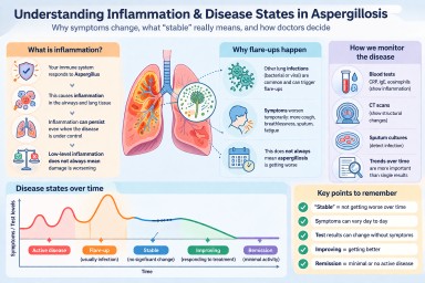
Inflammation and Aspergillosis: Understanding “Stable”, “Flare”, and “Improving” Disease
Last reviewed: April 2026
Key points
- Inflammation is part of the body’s response to Aspergillus, but it does not always mean damage is actively worsening.
- “Stable” disease means no clear progression over time, not that the condition has disappeared.
- Symptoms in aspergillosis often vary because of other infections, especially in the lungs.
- Test results (such as IgE or CRP) can change without symptoms changing.
- Doctors make decisions based on the overall pattern over time, not a single test result.
Table of contents
- What is inflammation and why does it matter?
- Inflammation in different types of aspergillosis
- Clear definitions: active, flare, stable, improving, remission
- What does “stable disease” mean in practice?
- Why other infections cause flare-ups
- Understanding test results (CRP, IgE, scans)
- When test results worsen but symptoms do not
- How doctors decide what is happening
- Common patient questions
- When to seek medical advice
What is inflammation and why does it matter?
Inflammation is the body’s way of responding to something it sees as harmful. In aspergillosis, this is usually the fungus Aspergillus.
This response involves immune cells, chemicals, and changes in the lungs that aim to control the fungus. However, if inflammation continues over a long period (chronic inflammation), it can also contribute to:
- Ongoing symptoms (cough, breathlessness, fatigue)
- Mucus production
- Damage to lung tissue over time
Important: inflammation can be present at a low level without causing active damage. This is common in chronic conditions.
Inflammation in different types of aspergillosis
The type of inflammation depends on the form of aspergillosis:
- Allergic Bronchopulmonary Aspergillosis (ABPA): driven by an overactive allergic response. Blood markers such as IgE and eosinophils are often used to monitor this.
- Chronic Pulmonary Aspergillosis (CPA): caused by long-term infection in damaged lung tissue, leading to ongoing inflammation and structural changes.
- Aspergillus bronchitis: persistent infection with inflammation, often causing chronic cough and sputum.
In all cases, inflammation may improve with treatment but often does not disappear completely.
Clear definitions: disease states
Doctors use the following terms to describe how the disease is behaving:
- Active disease: symptoms, tests, or scans are getting worse over time
- Flare-up: a short-term worsening, often triggered by infection or another stress on the body
- Stable: no clear overall change over time
- Improving / responding to treatment: symptoms and/or tests are getting better
- Remission: minimal or no signs of active disease (used more often in ABPA)
Key point: these states are not fixed — patients may move between them.
What does “stable disease” mean in practice?
“Stable” means that, over a period of time (weeks to months), there is no clear evidence that the disease is progressing.
This usually includes:
- No worsening of key symptoms
- No new complications (e.g. haemoptysis, significant weight loss)
- Imaging (CT scans) showing no progression
- No need to increase treatment
What stable does NOT mean:
- It does not mean symptoms are absent
- It does not mean inflammation is zero
- It does not mean you will feel the same every day
Many patients with stable disease still experience day-to-day variation in symptoms.
Why other infections cause flare-ups
People with aspergillosis are more vulnerable to other lung infections (bacterial or viral).
This is because:
- Lung structure may already be damaged
- Mucus clearance is less effective
- The immune system is already active
When another infection occurs, it can trigger a flare-up, causing:
- Increased cough and breathlessness
- More or thicker sputum
- Fatigue and feeling unwell
- Raised inflammatory markers (e.g. CRP)
Crucial point: this does not necessarily mean the aspergillosis itself is worsening. It is often a temporary additional problem.
Understanding test results
Doctors use several types of tests to monitor inflammation and disease activity:
- CRP / ESR: general markers of inflammation
- IgE: particularly important in ABPA
- Eosinophils: linked to allergic inflammation
- CT scans: show structural changes in the lungs
- Sputum cultures: detect infection
Important limitations:
- No single test gives a complete picture
- Results can fluctuate for many reasons
- Changes must be interpreted over time
When test results worsen but symptoms do not
This situation is common, especially in ABPA.
For example, IgE levels may rise without any noticeable change in symptoms.
This may happen because of:
- Natural biological variation
- Exposure to allergens
- A mild or early flare that has not yet caused symptoms
Key point: a change in a single test result does not automatically mean the disease is worsening.
Doctors will usually:
- Repeat tests
- Look for consistent trends
- Assess symptoms and scans
If symptoms remain stable and no other changes are seen, the condition may still be considered stable — but monitored more closely.
How doctors decide what is happening
Clinicians do not rely on a single result. Instead, they assess the pattern over time:
- Are symptoms changing?
- Are test results consistently rising or falling?
- Are scans stable or changing?
- Is the patient responding to treatment?
This combined assessment is called the clinical picture.
Common questions
If I feel better, what is that called?
This is usually described as improving or responding to treatment. In some cases (especially ABPA), it may be called remission.
Does inflammation always mean damage?
No. Low-level inflammation can persist without causing further harm.
Why do my symptoms change from day to day?
This is common and often relates to infections, environment, or general health rather than disease progression.
Can aspergillosis affect the whole body?
It can have wider effects, but it mainly affects the lungs in most patients.
When to seek medical advice
Seek medical advice if you notice:
- Persistent worsening of symptoms
- New haemoptysis (coughing up blood)
- Significant weight loss
- Symptoms not improving after a suspected infection
- Concerns about test results
Author and review
Author: Aspergillosis Patient Education Team
Reviewed by: National Aspergillosis Centre (UK)
References
- Denning DW et al. Chronic pulmonary aspergillosis guidelines
- ISHAM ABPA guidelines
This article is for general information only and is not a substitute for medical advice.
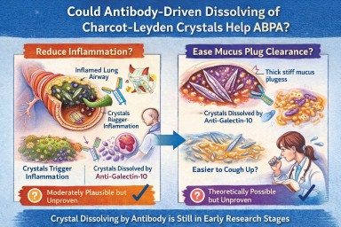
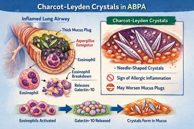
🧬 Could Antibody-Driven Dissolving of Charcot–Leyden Crystals Help ABPA?
Researchers have recently discovered that Charcot–Leyden crystals (CLCs) — the needle-shaped structures formed from the eosinophil protein galectin-10 — are not just debris.
In laboratory studies, specially designed antibodies can dissolve these crystals.
This has raised two important questions:
-
Could dissolving the crystals reduce airway inflammation?
-
Could dissolving them make mucus plugs easier to clear?
Here is what we currently know.
1️⃣ Could dissolving crystals reduce airway inflammation?
What we know
Laboratory and animal studies have shown:
-
Charcot–Leyden crystals can activate immune cells (especially macrophages).
-
They can stimulate inflammatory pathways (including inflammasome signalling).
-
In mouse models, antibodies targeting galectin-10 dissolved the crystals.
-
When crystals were dissolved, airway inflammation decreased.
This suggests that the crystals themselves may amplify inflammation, rather than simply mark it.
What this means biologically
In ABPA and eosinophilic asthma:
-
Eosinophils release galectin-10.
-
Galectin-10 crystallises.
-
Crystals may trigger further immune activation.
-
That leads to more inflammation → more eosinophils → more crystals.
Dissolving the crystals could theoretically interrupt this feedback loop.
How likely is this to help inflammation in humans?
Moderately plausible, but not yet proven.
The biological mechanism is strong.
The animal data are encouraging.
But no human clinical trials have yet shown reduced inflammation through crystal dissolution.
If developed successfully, this approach could:
-
Reduce airway immune activation
-
Lower exacerbation risk
-
Potentially reduce steroid dependence
But at present, it remains investigational.
2️⃣ Could dissolving crystals make mucus plugs easier to cough up?
This is more speculative — but still biologically reasonable.
Why mucus plugs are so thick in ABPA
ABPA mucus plugs contain:
-
Gel-forming mucins
-
DNA from inflammatory cells
-
Dead cells
-
Fungal fragments
-
Eosinophil proteins
-
Charcot–Leyden crystals
The crystals are:
-
Rigid
-
Needle-shaped
-
Structurally stable
When embedded in mucus, they likely increase:
-
Mechanical stiffness
-
Plug density
-
Resistance to deformation
From a physics perspective:
Removing rigid crystalline structures from a gel should reduce stiffness and improve flow.
Do we have direct evidence?
No.
There are currently:
-
No human studies measuring mucus clearance after crystal dissolution
-
No trials showing improved plug expectoration from crystal-targeting therapy
So while it is plausible that dissolving crystals could soften plugs, this has not yet been demonstrated in patients.
3️⃣ How strong is the overall case?
| Outcome | Evidence strength | Likelihood |
|---|---|---|
| Reduced inflammation | Strong biological rationale + animal data | Moderately promising |
| Easier mucus clearance | Biophysical plausibility only | Possible but unproven |
Inflammation reduction is the more evidence-supported target.
Improved plug clearance is plausible but currently theoretical.
4️⃣ How does this compare to existing treatments?
Current therapies (e.g., anti-IL-5 biologics) reduce eosinophils upstream.
That leads to:
-
Less galectin-10 release
-
Fewer crystals forming
-
Reduced inflammation
-
Often improved mucus plugging
So biologics already indirectly reduce crystal burden.
A crystal-dissolving antibody would act downstream, targeting the structural product directly.
This could theoretically:
-
Accelerate resolution of existing plugs
-
Reduce residual inflammatory signalling
But again, this remains in early research stages.
5️⃣ Practical take-home message
At present:
-
Dissolving Charcot–Leyden crystals reduces inflammation in animal models.
-
It is biologically plausible that this could also soften mucus plugs.
-
There is no human clinical proof yet.
-
No approved therapy currently targets the crystals directly.
The concept is scientifically credible — but still under development.
🔭 The Bigger Picture
ABPA is increasingly understood as a condition driven by:
-
Eosinophils
-
Allergic immune signalling
-
Abnormal mucus biology
-
Structural plug formation
Crystal-targeting therapies may eventually become part of a more precise approach to treating eosinophilic airway disease.
But for now, they remain a promising research direction rather than a clinical option.
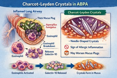
🔬 Charcot–Leyden Crystals in ABPA and Asthma
What are they? Why do they form? Do they matter?
If you live with Allergic Bronchopulmonary Aspergillosis (ABPA) or severe asthma, you may see the term Charcot–Leyden crystals in a sputum or pathology report.
They can sound worrying.
They are:
-
Not fungus
-
Not infection
-
Not cancer
They are a sign of a particular type of allergic inflammation in the airways.
🧬 What Are Charcot–Leyden Crystals?
Charcot–Leyden crystals are microscopic, needle-shaped structures found in mucus.
They are made from a protein called galectin-10, which is stored inside a type of white blood cell called an eosinophil.
Eosinophils are immune cells involved in:
-
Allergic asthma
-
ABPA
-
Severe asthma with fungal sensitisation
-
Parasitic infections
When eosinophils are activated and break down, they release galectin-10.
If enough of this protein accumulates in thick airway mucus, it crystallises into visible crystals.
So the crystals are made from your immune cells, not from Aspergillus.
🫁 Why Do They Appear in ABPA?
In ABPA:
-
The immune system overreacts to Aspergillus fumigatus.
-
This triggers a strong allergic (Type 2) immune response.
-
Large numbers of eosinophils move into the airways.
-
Eosinophils break down and release galectin-10.
-
The protein crystallises inside mucus plugs.
The crystals are therefore a footprint of intense allergic inflammation, not fungal invasion.
🌡 Is Most ABPA Eosinophilic?
Yes — almost all classical ABPA is eosinophilic.
ABPA is fundamentally a Type 2 allergic condition, driven by immune pathways involving:
-
IL-4
-
IL-5
-
IL-13
-
IgE
-
Eosinophils
IL-5 in particular stimulates eosinophil production and survival.
Because of this, eosinophils are central to the disease process.
Historically, raised blood eosinophils have been part of diagnostic criteria.
However:
-
Eosinophil counts can fluctuate
-
Steroids can suppress blood levels
-
Eosinophils may still be present in airway mucus even if blood counts appear normal
So ABPA is biologically eosinophilic — even if a single blood test does not show a high count.
True non-eosinophilic ABPA would be unusual and would prompt clinicians to reconsider the diagnosis.
❓ Are Crystals Caused by Aspergillus Infection?
No.
They are caused by the immune reaction to Aspergillus — not by the fungus itself.
They can also be seen in:
-
Severe eosinophilic asthma
-
Parasitic infections
-
Other allergic lung conditions
They reflect eosinophil activity, not fungal growth.
🧠 Why Don’t All People with Asthma Develop These Crystals?
Asthma is not one single disease. It has different inflammatory patterns.
Type 2 (Eosinophilic) Asthma
This involves high eosinophils and allergic pathways.
Common in:
-
Allergic asthma
-
ABPA
-
Severe eosinophilic asthma
These patients can develop Charcot–Leyden crystals.
Non–Type 2 (Non-Eosinophilic) Asthma
This includes:
Neutrophilic asthma
Driven by neutrophils rather than eosinophils.
Paucigranulocytic asthma
Very few inflammatory cells present.
In these forms:
-
Eosinophils are low
-
Galectin-10 is not released in large amounts
-
Crystals are unlikely to form
🧱 Do Charcot–Leyden Crystals Make Mucus Plugs Worse?
Possibly.
Research suggests they may:
-
Increase mucus thickness
-
Contribute mechanically to airway blockage
-
Stimulate further inflammation
For many years they were thought to be harmless debris.
Modern studies suggest they may actively amplify inflammation when present in large amounts.
🎯 Do They Have a Purpose?
Eosinophils evolved mainly to help fight parasitic infections.
Galectin-10 probably has immune signalling roles inside cells.
However, when large amounts are released into thick airway mucus, crystallisation appears to be a by-product of excessive immune activity rather than a useful defence.
In ABPA and allergic asthma, they are more likely part of the problem than part of the solution.
💧 Can Their Formation Be Reduced?
Hydration alone does not stop them forming.
Drinking fluids helps:
-
Keep mucus less sticky
-
Support airway clearance
But it does not prevent eosinophils releasing galectin-10.
What reduces crystal formation?
Reducing eosinophilic inflammation:
-
Corticosteroids
-
Anti-IL-5 biologics
-
Anti-IL-4/IL-13 biologics
When eosinophil numbers fall:
→ Less galectin-10 is released
→ Fewer crystals form
Antifungal treatment in ABPA may indirectly help by reducing allergic stimulation, but the main driver is the immune response.
📊 Do They Change Treatment?
Not directly.
Doctors base treatment on:
-
Symptoms
-
Blood eosinophils
-
Total IgE
-
Imaging
-
Lung function
-
Exacerbation history
Crystals support the diagnosis of eosinophilic inflammation but do not determine treatment alone.
🔎 What Do They Tell Us?
Charcot–Leyden crystals tell us:
-
The airway inflammation is eosinophilic.
-
The immune response is strongly allergic.
-
Mucus plugging risk may be higher.
They are a marker of immune overreaction, not infection severity.
🧠 Key Points to Remember
-
They are made from proteins released by eosinophils.
-
They are not Aspergillus.
-
They do not mean invasive fungal infection.
-
Most classical ABPA is eosinophilic.
-
They are unlikely in non-eosinophilic asthma.
-
Reducing eosinophils reduces their formation.
-
Hydration helps clearance but does not prevent formation.
In simple terms:
Charcot–Leyden crystals are microscopic signs that the immune system is working too hard in the airways.
Aspergillosis, immunity, and risk
Primary immune deficiencies and immune modifiers explained
A single, comprehensive explainer for expert patients, carers, and non-specialists
Why this article exists
Aspergillus is a mould that everyone breathes in every day. Most people clear it without difficulty.
A small number of people develop aspergillosis because the balance between the fungus, the lungs, and the immune system is disturbed.
This article explains:
-
Rare primary (inherited) immune deficiencies that are clearly linked to aspergillosis
-
Common immune “modifier” factors that can increase risk or severity but do not cause disease on their own
-
How these factors stack together in real life
Key reassurance up front
There are 500+ recognised primary immune deficiencies
Only ~20–30 are clearly linked to aspergillosis
Most people with aspergillosis do not have any inherited immune disorder
The unifying concept: three immune pathways to aspergillosis
Almost all immune–aspergillus relationships fall into three mechanisms. Understanding these matters more than memorising names.
1. Reduced ability to kill the fungus
Some immune cells fail to destroy Aspergillus spores effectively.
→ Risk of invasive aspergillosis, sometimes severe or life-threatening.
2. Lung damage over time
Repeated infections or inflammation damage airways or leave cavities.
→ Risk of chronic pulmonary aspergillosis (CPA) or aspergillomas.
3. Excessive allergic inflammation
The immune system over-reacts to Aspergillus rather than failing to fight it.
→ Allergic bronchopulmonary aspergillosis (ABPA) and severe fungal-sensitised asthma.
Many conditions overlap more than one pathway.
Section 1: Primary (inherited) immune deficiencies clearly linked to aspergillosis
Rare, high-impact, and sometimes life-changing when present
These are the conditions clinicians usually mean when they talk about “immune causes of aspergillus disease”.
A. Phagocyte defects
Strongest association with invasive aspergillosis
-
Chronic granulomatous disease (CGD)
-
Autosomal recessive forms of CGD
-
Severe congenital neutropenia
-
Cyclic neutropenia
-
Leukocyte adhesion deficiency type I
Typical pattern
-
Aspergillosis at a young age
-
Invasive lung disease ± spread beyond lungs
-
Often no other obvious risk factors
B. Hyper-IgE and severe allergy syndromes
Allergic, chronic, and cavity-associated disease
-
STAT3 hyper-IgE syndrome
-
DOCK8 deficiency
-
PGM3 deficiency
-
ZNF341 deficiency
-
IL6ST deficiency
Typical pattern
-
Severe asthma and allergy
-
Thick mucus, recurrent infections
-
ABPA, later CPA or aspergillomas
C. Combined immunodeficiencies
Immune coordination problems
-
Severe combined immunodeficiency (milder or surviving forms)
-
Omenn syndrome
-
ZAP-70 deficiency
-
Major histocompatibility complex class II deficiency
-
CD40 ligand deficiency (hyper-IgM syndrome)
Typical pattern
-
Broad infection susceptibility
-
Aspergillosis can behave aggressively
D. Defects of fungal recognition and innate signalling
Often dramatic or unexpected presentations
-
CARD9 deficiency
-
Dectin-1 (CLEC7A) complete deficiency
-
MALT1 deficiency
Typical pattern
-
Severe or unusual aspergillosis
-
Lung, brain, or deep tissue involvement
-
Sometimes first presents in adulthood
E. Immune dysregulation syndromes
Mixed infection, inflammation, and autoimmunity
-
CTLA-4 haploinsufficiency
-
LRBA deficiency
-
STAT1 gain-of-function mutations
-
IPEX syndrome (FOXP3 deficiency)
Typical pattern
-
Inflammatory lung disease
-
Chronic or invasive aspergillosis emerging over time
F. Antibody deficiencies (indirect risk via lung damage)
-
Common variable immunodeficiency
-
X-linked agammaglobulinaemia
-
Activated PI3K-delta syndrome
Important nuance
Antibodies do not normally kill Aspergillus.
Risk arises after years of lung damage, not early in life.
Section 2: Immune modifier-types that can amplify risk
Common, low-penetrance, and often invisible on routine testing
These are not immune deficiencies, but they can influence who struggles, how severely, and why disease persists.
Mannose-binding lectin (MBL) deficiency
-
Common (≈5–10% of population)
-
Affects fungal recognition and complement activation
-
Usually mild on its own
-
Becomes relevant with lung disease, steroids, or other immune issues
Partial fungal-recognition receptor variants
-
Heterozygous dectin-1 variants
-
Toll-like receptor polymorphisms (for example TLR2, TLR4)
Effect
-
Slower fungal recognition
-
Increased colonisation or allergic response
-
Act as risk amplifiers, not causes
Cytokine balance variants
Small genetic differences affecting immune “signal strength”, including:
-
Interleukin-6
-
Interleukin-10
-
Tumour necrosis factor-alpha
These modify:
-
Inflammation intensity
-
Tissue damage vs clearance balance
Allergy-biased (Th2-skewed) immunity
Not a disease, but a recognised immune tendency.
Features:
-
Asthma
-
Eczema
-
Nasal polyps
-
High immunoglobulin E levels
-
Eosinophilia
Strongly associated with:
-
Fungal sensitisation
-
ABPA
-
Difficult-to-control asthma
Impaired mucociliary clearance
A functional immune–mechanical issue.
Seen in:
-
Severe asthma
-
Bronchiectasis
-
Chronic sinus disease
Effect:
-
Aspergillus spores are not physically cleared
-
Prolonged immune exposure
-
Increased colonisation and allergy
Age-related immune change (immunosenescence)
-
Normal reduction in immune speed and coordination with age
-
Particularly relevant to chronic pulmonary aspergillosis
Not a disease, but an important modifier of outcome.
Airway epithelial vulnerability
Subtle weaknesses in:
-
Airway lining integrity
-
Antimicrobial peptide production
-
Local immune signalling
Can increase:
-
Fungal adherence
-
Chronic colonisation
-
Allergic sensitisation
Section 3: Risk stacking – how this works in real life
Aspergillosis rarely results from one single factor.
Instead, several modest risks align:
-
Mild MBL deficiency
-
Severe asthma
-
Corticosteroid exposure
-
Bronchiectasis
-
Age-related immune change
→ Together, they create real disease risk, even though none alone would.
This explains why:
-
Two people with similar scans can behave very differently
-
One patient relapses while another stabilises
-
“Why me?” often has no single answer
Section 4: When clinicians investigate immune causes
Testing is targeted, not routine. It is usually considered when there is:
-
Aspergillosis at a young age
-
Invasive or unusually severe disease
-
Disease without classic risk factors
-
Recurrent infections plus severe asthma or allergy
-
A family history of unusual infections
Section 5: Why identifying (or excluding) immune factors helps
Understanding immune contribution can:
-
Explain disease pattern and behaviour
-
Guide antifungal choice and duration
-
Inform long-term prevention strategies
-
Reduce future lung damage
-
Reassure patients when no immune defect is found
Key take-home messages
-
Aspergillus exposure is universal; immune causes are rare
-
Only ~20–30 inherited immune deficiencies are clearly linked to aspergillosis
-
Modifier-type immune factors are common and usually harmless alone
-
Aspergillosis often reflects risk stacking, not a single diagnosis
-
Understanding patterns matters more than labels
-
Specialist care improves precision and outcomes
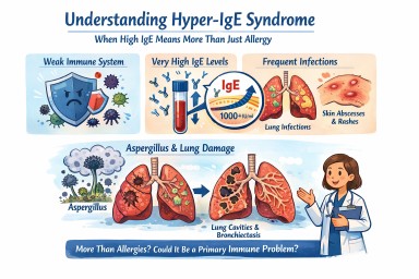
Hyper-IgE syndrome
A patient-friendly guide (and why it matters if you have aspergillosis)
It is not the same as having lots of allergies, even though it can look very similar at first.
What is IgE, and why does it matter?
IgE is usually involved in allergies and asthma.
In Hyper-IgE syndrome:
-
IgE levels are extremely high (often many thousands)
-
But the immune system is unbalanced
-
This makes infections—especially in the lungs and skin—harder to control
So IgE is high, but protection is weak.
How might Hyper-IgE syndrome affect everyday life?
Not everyone has the same symptoms, but common features include:
Lung and chest problems
-
Repeated chest infections (often from a young age)
-
Ongoing cough, breathlessness and mucus
-
Lung damage such as bronchiectasis
-
Lung cavities that can later become infected by moulds such as Aspergillus
Skin and infection problems
-
Long-standing eczema or very sensitive skin
-
Recurrent skin infections or boils
-
Infections that keep coming back or take a long time to clear
Other clues (in some people)
-
Frequent infections in childhood
-
Bone or joint problems
-
Dental issues (for example baby teeth not falling out on time)
Why is this important for people with aspergillosis?
For many people, Aspergillus causes allergy or irritation.
In Hyper-IgE syndrome:
-
The immune system struggles to control moulds
-
Aspergillus can behave more like a true infection, not just an allergy
-
Lung damage can happen more easily and progress faster
This means doctors may need to:
-
Monitor lungs more closely
-
Treat fungal disease earlier and for longer
-
Be cautious with repeated or long-term steroid use
Specialist centres such as the National Aspergillosis Centre are often involved when aspergillosis and immune problems overlap.
Isn’t this just severe allergy or ABPA?
Hyper-IgE syndrome can look similar to:
-
Severe allergic asthma
-
Allergic Bronchopulmonary Aspergillosis (ABPA)
The key difference is that in Hyper-IgE syndrome:
-
The immune system itself is faulty
-
High IgE is part of a wider immune problem
-
Treating allergy alone may not be enough
Some people are treated for asthma or ABPA for years before this possibility is considered.
How is Hyper-IgE syndrome treated?
There is no single cure, but good treatment can make a big difference. The aim is to prevent infections, protect the lungs, and reduce symptoms.
1. Preventing infections (most important)
Because the immune system does not fight germs normally:
-
Some people take regular low-dose antibiotics
-
Others use antibiotics early and promptly when infections start
For people with aspergillosis:
-
Antifungal medicines may be needed
-
Monitoring is usually closer and longer-term
2. Protecting the lungs
Many people develop bronchiectasis or lung damage, so care often includes:
-
Airway clearance physiotherapy
-
Saline nebulisers to help clear mucus
-
Regular sputum tests
-
Early treatment of flare-ups
The goal is to stop the cycle of:
infection → inflammation → permanent lung damage
3. Managing inflammation and allergy (carefully)
People may also have asthma-like symptoms, eczema and multiple allergies.
-
Steroids can help symptoms, but long-term or frequent use can increase infection risk
-
Doctors usually try to keep steroid doses as low as possible
Biologic treatments (such as anti-IgE medicines):
-
May help some people
-
Do not fix the immune problem
-
Are considered on an individual basis, usually in specialist centres
4. Skin care
-
Regular moisturising
-
Prompt treatment of infected eczema
-
Good skin care helps reduce infection risk
How is Hyper-IgE syndrome diagnosed?
Diagnosis usually involves:
-
A detailed review of your medical history (often including childhood infections)
-
Blood tests of immune function
-
Referral to an immunology specialist
-
Sometimes genetic testing
Does having high IgE mean I definitely have this?
No.
Hyper-IgE syndrome is rare.
But it may be worth asking about if:
-
Your IgE has always been extremely high
-
You’ve had repeated infections for many years
-
You have bronchiectasis without a clear cause
-
Aspergillosis seems unusually persistent or severe
-
Standard asthma or allergy treatments don’t fully explain your symptoms
Key message
Very high IgE does not always mean “just allergy.”
In a small number of people, it reflects a deeper immune problem that changes how aspergillosis behaves and how it should be treated.
If your illness doesn’t quite fit the usual labels, it is reasonable to ask whether an immunology review would help.

**Understanding Your Immune System:
A Simple Guide for Aspergillillosis Patients and Carers**
Part of the Aspergillosis Immune System Knowledge Hub
(See also: Articles 2, 3, and 4)
When you live with aspergillosis, asthma, bronchiectasis, or fungal allergy, the immune system plays a major role in your symptoms and how your condition behaves. This article explains the key parts of the immune system in a clear, accessible way.
🧬 1. B Cells — the Antibody Makers
B cells produce antibodies, which act like “tags” that help the immune system recognise germs.
They make different types, including:
-
IgE — triggers allergy
-
IgG — provides long-term immunity and helps diagnose chronic infection
-
IgA — protects the nose, throat, and gut
In Aspergillosis:
-
ABPA: B cells overproduce IgE against Aspergillus.
-
CPA: High Aspergillus IgG helps confirm chronic infection.
-
SAFS/Bronchitis: Mixed or subtle antibody patterns.
🧠 2. T Cells — the Immune System’s Directors
T cells guide and regulate the immune response.
Types include:
-
Helper T cells (Th cells): tell B cells what antibodies to make
-
Killer T cells: destroy infected or damaged cells
-
Regulatory T cells: calm the immune system and prevent over-reaction
In Aspergillosis:
-
ABPA: Helper T cells become overactive, driving allergic inflammation.
-
CPA: T cells attempt to control fungal growth but cannot fully clear it.
🟡 3. IgE — the Allergy Antibody
IgE causes:
-
wheezing
-
swelling
-
itching
-
mucus production
-
allergic reactions
In ABPA, IgE levels become very high because the body incorrectly treats Aspergillus as a major allergen.
🟢 4. IgG — the Memory and Detection Antibody
IgG helps the immune system remember past infections.
A raised Aspergillus IgG level is one of the main tests for CPA.
🔥 5. Mast Cells — the Alarm Cells
Mast cells sit in the lungs, nose, sinuses, skin, and gut.
When triggered (often by IgE), they release:
-
histamine
-
leukotrienes
-
inflammatory chemicals
This causes:
-
wheezing
-
chest tightness
-
mucus production
-
itching or burning sensations
-
coughing
They are very active in ABPA and severe asthma.
🌈 6. Histamine — Why Symptoms Feel the Way They Do
Histamine release leads to:
-
swelling and redness
-
increased mucus
-
nerve irritation → itch, tickle, burning
-
airway narrowing → wheeze and breathlessness
This explains why flare-ups can feel sudden or “out of proportion” to test results.
🧩 7. Putting It Together: Immune Pathways in Aspergillosis
| Condition | Dominant Antibody | Key Cells | Symptoms Driven By |
|---|---|---|---|
| ABPA | Very high IgE | Mast cells, eosinophils | Allergy, mucus, flare-ups |
| CPA | Raised IgG | T cells, macrophages | Chronic inflammation, cavities |
| SAFS/Allergic asthma | IgE ± eosinophils | Mast cells, eosinophils | Wheeze, mucus, sensitivity |
| Aspergillus bronchitis | Variable | Neutrophils, airway cells | Cough, sputum, recurring infections |
Understanding these pathways helps you and your clinical team choose the right treatments.
Next articles:
- **Understanding your immune system
- How the Immune System Knows “Self”
- What Happens in Autoimmune Disease (Addison’s Explained)
- Eosinophils and Type-2 Inflammation in Aspergillosis
- **Where Do All These Immune Cells Live, and Where Are They Made?

**Where Do All These Immune Cells Live, and Where Are They Made?
A Simple Guide for Patients and Carers**
When we talk about T cells, B cells, eosinophils, mast cells, IgE, IgG, and other immune system parts, it’s natural to wonder:
Where are these cells actually made?
Where do they live in the body?
Where do they go when you’re ill?
Here is a simple explanation.
🧱 1. Most immune cells are MADE in the bone marrow
Bone marrow is the soft tissue inside your bones (especially the pelvis, spine, ribs, skull, and sternum).
Inside this marrow are stem cells, which are the “mother cells” that can turn into:
-
red blood cells
-
white blood cells
-
platelets
Almost all immune cells begin their life in the bone marrow, including:
-
B cells
-
eosinophils
-
mast cell precursors
-
neutrophils
-
monocytes
-
basophils
The bone marrow is like the main factory for your entire immune system.
🫀 2. T cells are trained in the thymus
After T cells are created in the bone marrow, they travel to the thymus — a small organ behind the breastbone.
The thymus is like a school where T cells learn:
-
what is safe
-
what is dangerous
-
how to avoid attacking the body itself
This training is essential for preventing autoimmune diseases.
After training, T cells spread through the body.
🩸 3. Immune cells travel in the blood and lymph
Once made, immune cells circulate around the body like security guards on patrol.
They travel through:
Blood
This carries cells quickly to any part of the body.
Lymph system
A drainage and communication network that runs alongside the bloodstream.
Lymph nodes (in the neck, armpits, groin) act like checkpoints, where:
-
immune cells meet
-
information is exchanged
-
inflammation signals get amplified
If your glands are swollen during illness, that’s because immune cells are gathering there.
🫁 4. Many immune cells live in tissues, not just in the blood
Some immune cells settle in certain places:
Mast cells
Live in tissues such as:
-
lungs
-
sinuses
-
skin
-
gut
-
blood vessels
They wait there like "alarm sensors," ready to react if something enters the tissue.
Macrophages
Live in tissues and “eat” germs.
Eosinophils
Move into tissues during allergy or asthma flare-ups.
T cells and B cells
Live in:
-
lymph nodes
-
spleen
-
tonsils
-
tissues throughout the body
-
airway lining in people with asthma or ABPA
🧫 5. Where antibodies (IgE, IgG) come from
Antibodies are made by plasma cells, which are specialised B cells.
These plasma cells usually live in:
-
the bone marrow
-
lymph nodes
-
spleen
-
airway tissues (especially in chronic inflammation)
So:
-
IgE is mostly made in tissues involved in allergy (lungs, sinuses, skin).
-
IgG is made in bone marrow and lymph tissues to provide long-term protection.
Antibodies then circulate in the blood, ready to recognise anything they have been trained to detect.
🧬 Where these cells actually are, in simple terms:
| Immune Cell / Antibody | Where It Is Made | Where It Lives / Works |
|---|---|---|
| B cells | Bone marrow | Lymph nodes, blood, tissues |
| Plasma cells (make antibodies) | Bone marrow / lymph nodes | Bone marrow, tissues |
| T cells | Bone marrow → trained in thymus | Blood, lymph nodes, organs |
| IgE antibodies | Plasma cells in tissues | Lungs, skin, blood |
| IgG antibodies | Plasma cells | Blood (body-wide protection) |
| Eosinophils | Bone marrow | Blood → lungs during flare-ups |
| Mast cells | Bone marrow (as precursors) | Lungs, skin, sinuses, gut |
| Neutrophils | Bone marrow | Blood → infection sites |
🧠 6. How this applies to aspergillosis
In ABPA
-
IgE is made in the lung tissues.
-
Mast cells in the lungs release histamine.
-
Eosinophils move from the bone marrow into the airways.
In CPA
-
IgG is made in bone marrow in response to chronic infection.
-
T cells gather in lung cavities and damaged tissue.
In fungal asthma / SAFS
-
Mast cells and eosinophils in the lungs respond strongly to triggers.
Understanding where these cells come from and where they live helps explain why:
-
symptoms can flare suddenly
-
blood test levels change
-
treatments like steroids or biologics work
-
inflammation can persist even when scans look stable
🏁 Simple takeaway
-
Your bone marrow makes most of your immune cells.
-
Your thymus trains T cells.
-
Immune cells patrol your blood and lymph system.
-
Many immune cells live long-term in your lungs, skin, and tissues.
-
Antibodies are made by plasma cells in bone marrow and lymph nodes.
-
In aspergillosis, the lungs become a major “immune battlefield.”
Next articles:
- **Understanding your immune system
- How the Immune System Knows “Self”
- What Happens in Autoimmune Disease (Addison’s Explained)
- Eosinophils and Type-2 Inflammation in Aspergillosis
- **Where Do All These Immune Cells Live, and Where Are They Made?

**Eosinophils and Type-2 Inflammation:
What Aspergillosis Patients Need to Know**
Part of the Aspergillosis Immune System Knowledge Hub
Eosinophils are a type of white blood cell central to allergy, asthma, and ABPA. They play a major role in symptoms, flare-ups, mucus plugging, and treatment responses.
This article explains eosinophils in simple terms.
🧬 1. What Are Eosinophils?
Eosinophils are immune cells filled with granules containing powerful enzymes.
They normally help:
-
fight parasites
-
regulate allergic inflammation
-
repair tissues
-
produce important immune signals
But in excess, they can cause damage — especially in the lungs.
🔥 2. Eosinophils in the Lungs
Activated eosinophils release their granules into airway tissues, causing:
-
swelling
-
increased mucus
-
airway narrowing
-
cough sensitivity
-
wheezing
-
breathlessness
This makes them key players in allergic and fungal-related lung disease.
🌟 3. Eosinophils in ABPA
Eosinophils are highly active in ABPA.
ABPA involves a strong “type-2” allergic response to Aspergillus, including:
-
high IgE
-
mast cell activation
-
large numbers of eosinophils
-
thick, sticky mucus
-
airway obstruction
-
repeated flare-ups
Eosinophils contribute significantly to long-term lung damage if not controlled.
🌬 4. Eosinophils in Severe Asthma and SAFS
In severe or allergic asthma:
-
eosinophils can be persistently high
-
they drive airway swelling
-
they increase sensitivity to triggers
-
they worsen recovery after infection
In SAFS, eosinophils may be moderately raised but symptoms can still be severe.
🦠 5. Eosinophils in CPA
In CPA, eosinophils are not usually the dominant cell, but they still matter when patients also have:
-
asthma
-
ABPA overlap
-
fungal allergy
-
airway hypersensitivity
-
steroid withdrawal flare-ups
🔗 6. How Eosinophils Link to Other Immune Cells
They interact with:
-
IgE → recruits eosinophils
-
T-helper cells (Th2) → tell bone marrow to make more
-
Mast cells → release histamine that pulls eosinophils into tissues
-
Airway lining cells → release distress signals
This is why severe allergic pathways often involve all three:
IgE → mast cells → eosinophils
💊 7. Treatments That Target Eosinophils
✔ Steroids (oral or inhaled)
Suppress eosinophil activity.
✔ Biologics
Directly reduce eosinophils:
-
Mepolizumab (anti-IL-5)
-
Benralizumab (anti-IL-5 receptor)
-
Reslizumab (anti-IL-5 infusion)
Reduce eosinophil recruitment:
-
Dupilumab (anti-IL-4/IL-13)
-
Tezepelumab (broad upstream suppression)
These can transform life for patients with severe asthma or ABPA.
🧠 8. Summary
Eosinophils are key drivers of:
-
flare-ups
-
mucus plugging
-
wheeze
-
breathlessness
-
airway damage
Understanding them helps patients:
-
interpret blood tests
-
understand biologic treatments
-
recognise flare-up patterns
-
manage ABPA and asthma more confidently
Next articles:
- **Understanding your immune system
- How the Immune System Knows “Self”
- What Happens in Autoimmune Disease (Addison’s Explained)
- Eosinophils and Type-2 Inflammation in Aspergillosis
- **Where Do All These Immune Cells Live, and Where Are They Made?

**What Happens in Autoimmune Disease?
(Explained with Addison’s Disease)**
Part of the Aspergillosis Immune System Knowledge Hub
Autoimmune disease occurs when the immune system mistakenly attacks the body’s own tissues. This is different from allergy, infection, or inflammation caused by fungal disease. Addison’s disease is a clear example of autoimmunity and helps explain how this process works.
❌ 1. Autoimmunity = Loss of Immune Tolerance
In autoimmune disease:
-
the immune system starts recognising the body’s own tissues as “foreign”
-
T cells and B cells become misdirected
-
autoantibodies form
-
inflammation destroys healthy cells
This process develops over months or years.
🧬 2. What Specifically Happens in Addison’s Disease?
Addison’s disease is caused by an autoimmune attack on the adrenal cortex, the part of the adrenal gland that makes:
-
cortisol
-
aldosterone
-
DHEA (adrenal androgens)
The steps include:
1. Loss of tolerance
The immune system mistakenly targets adrenal enzymes (especially 21-hydroxylase).
2. Autoantibodies form
These can be detected in blood tests.
3. Cytotoxic T cells attack adrenal tissue
Gradually destroying hormone-producing cells.
4. Hormone levels fall
Leading to:
-
severe fatigue
-
weight loss
-
low blood pressure
-
salt loss
-
nausea
-
risk of adrenal crisis
Addison’s must be treated with lifelong hormone replacement.
🔄 3. Why Does Autoimmunity Happen?
Factors include:
✔ Genetic susceptibility (HLA types)
✔ Prior viral infection or severe inflammation
✔ Stressful life events
✔ Regulatory T-cell failure
✔ Microbiome disruption
✔ Hormonal influences
Importantly:
Autoimmune disease is never the patient’s fault.
🆚 4. How Autoimmunity Differs from Aspergillosis
| Aspergillosis | Autoimmune Disease |
|---|---|
| Driven by external organism (Aspergillus fungus) | Driven by immune system attacking “self” |
| ABPA → IgE allergies; CPA → IgG infection response | Autoantibodies + T-cell attack |
| Treatment aims at fungus + inflammation | Treatment replaces missing hormones |
| Damage = collateral | Damage = direct |
Some patients live with both conditions (e.g., ABPA + adrenal insufficiency), but they arise via very different mechanisms.
🧠 5. Key Message
Autoimmune disease results from a failure of immune tolerance, not from weakness, lifestyle, or exposure. Understanding this helps patients feel more in control and reduces self-blame.
Next articles:
- **Understanding your immune system
- How the Immune System Knows “Self”
- What Happens in Autoimmune Disease (Addison’s Explained)
- Eosinophils and Type-2 Inflammation in Aspergillosis
- **Where Do All These Immune Cells Live, and Where Are They Made?