
Artificial Intelligence (AI) in the NHS: Promise, Progress and the Future of Healthcare

Artificial intelligence (AI) is no longer a futuristic idea within the NHS. AI technologies are already being used in some areas of healthcare across the UK, particularly in radiology, diagnostics, administration and workflow support. However, the reality is more nuanced than many headlines suggest.
At present, AI in the NHS is best understood as a collection of promising technologies being introduced gradually, cautiously and under clinical oversight. Some NHS trusts are using several AI-assisted systems routinely, while others have little or no operational AI deployment beyond pilot projects.
The direction of travel is clear: AI is likely to become an increasingly important part of healthcare over the next decade. However, the NHS is moving carefully because healthcare is a high-risk environment where mistakes can have serious consequences.
What Does AI Mean in Healthcare?
In healthcare, AI usually refers to computer systems that can analyse large amounts of data and identify patterns more quickly or consistently than humans alone.
Examples include:
- Analysing medical scans
- Helping detect cancers or fractures
- Supporting diagnosis
- Summarising clinic consultations
- Reducing paperwork
- Predicting patient deterioration
- Helping prioritise urgent cases
- Supporting medication safety checks
Importantly, most NHS AI systems are currently designed to support clinicians rather than replace them.
AI in NHS Radiology
Radiology is currently one of the biggest areas of AI development within the NHS.
AI systems are being used or trialled to help analyse:
- Chest X-rays
- CT scans
- MRI scans
- Mammograms
- Retinal photographs
- Dermatology images
AI is particularly attractive in radiology because the NHS faces:
- Large radiologist shortages
- Increasing imaging demand
- Growing reporting backlogs
- Rising complexity of scans
How AI Is Currently Used
Most NHS radiology AI systems currently operate in one of three ways:
1. AI as a “Second Reader”
The scan is interpreted by a radiologist, while AI acts as an additional safety check.
AI may flag:
- Possible lung nodules
- Fractures
- Brain bleeds
- Abnormal mammograms
- Signs of stroke
The human clinician still makes the final decision.
2. Prioritisation (“Triage”)
AI can rapidly review scans and move potentially urgent cases higher in the reporting queue.
This may help speed up treatment for:
- Stroke
- Pulmonary embolism
- Pneumothorax (collapsed lung)
- Intracranial bleeding
3. Identifying Likely Normal Scans
Some AI systems are being studied to identify scans that appear very likely to be normal.
The aim is to allow radiologists to focus more attention on:
- Abnormal scans
- Complex cases
- Uncertain findings
However, this remains an area of careful evaluation and regulation.
Does AI Sometimes Perform Better Than Humans?
In certain narrow and repetitive tasks, AI can sometimes outperform humans or help reduce specific types of errors.
AI can be very good at:
- Detecting tiny abnormalities
- Maintaining consistency
- Working without fatigue
- Rapid image analysis
- Comparing huge numbers of images
Humans can become:
- Tired
- Distracted
- Overloaded
- Inconsistent under pressure
For example, AI may detect very small lung nodules or subtle fractures that could potentially be overlooked during a busy reporting session.
However, AI also has important weaknesses.
AI may struggle with:
- Rare diseases
- Complex clinical context
- Unusual anatomy
- Poor-quality scans
- Unexpected combinations of disease
Importantly, AI and humans often make different kinds of mistakes.
The emerging evidence suggests that:
Human + AI is often better than either alone.
Will Radiologists Be Replaced?
At present, the NHS does not view AI as a replacement for radiologists.
Instead, AI is mainly being introduced as:
- A support tool
- A safety net
- A prioritisation system
- A workflow assistant
Radiologists still provide:
- Clinical judgement
- Contextual interpretation
- Decision-making
- Communication
- Management of uncertainty
In the foreseeable future, radiology is likely to become increasingly AI-assisted but still human-led.
Longitudinal Analysis: One of the Most Exciting Future Possibilities
One of the most promising future applications of AI is longitudinal analysis.
This means comparing:
- Current scans
- Previous scans
- Blood tests
- Lung function
- Medications
- Clinical notes
- Symptoms
- Outcomes over time
Humans are not particularly good at consistently recognising very subtle changes across years of imaging and clinical data.
AI could potentially become extremely powerful at:
- Tracking disease progression
- Measuring tumour growth
- Monitoring fibrosis
- Quantifying cavity enlargement
- Identifying treatment response
- Predicting future deterioration
This could be especially valuable in chronic diseases such as:
- Chronic Pulmonary Aspergillosis (CPA)
- Bronchiectasis
- Chronic Obstructive Pulmonary Disease (COPD)
- Interstitial lung disease
- Cancer
In the future, AI may help move medicine from:
“What does this scan show today?”
towards:
“What is happening over time, and what is likely to happen next?”
AI Beyond Radiology
AI Clinical Documentation
The NHS is increasingly exploring AI systems that can generate:
- Clinic letters
- Consultation summaries
- Medical notes
- Coding suggestions
These “AI scribes” may help reduce administrative burden and allow clinicians to spend more time with patients.
AI Triage Systems
Some NHS services now use AI-assisted triage systems to:
- Route patient requests
- Identify urgent problems
- Prioritise appointments
- Support NHS App workflows
Medication Safety
AI may eventually help identify:
- Drug interactions
- Prescribing errors
- Missed monitoring
- Unsafe medication combinations
Operational Efficiency
The NHS is also exploring AI for:
- Appointment scheduling
- Referral management
- Staff rostering
- Reducing missed appointments
- Managing workflow
Why AI Adoption Is Uneven Across the NHS
AI adoption currently varies considerably across the NHS.
Some trusts use multiple AI systems routinely, while others have minimal deployment.
This variation is influenced by:
- Funding differences
- IT infrastructure
- Digital maturity
- Research partnerships
- Clinical confidence
- Procurement complexity
- Availability of evidence
Large teaching hospitals and academic centres often adopt new technologies earlier than smaller hospitals.
As a result, current NHS AI deployment is best described as:
Selective, cautious and evolving.
Why the NHS Is Proceeding Carefully
The NHS is naturally cautious about AI because healthcare is fundamentally different from many other industries.
Mistakes can have serious consequences, including:
- Missed cancers
- Delayed diagnosis
- Medication harm
- Unsafe treatment decisions
For this reason, NHS AI systems generally require:
- Clinical validation
- Governance review
- Safety monitoring
- Regulatory approval
- Human oversight
- Ongoing audit
There is also awareness that:
- commercial hype can exceed evidence,
- real-world NHS workflows are complex,
- and some AI systems may not perform as well outside carefully controlled studies.
Potential Risks and Concerns
Although AI has enormous potential, there are also important concerns.
Patient Safety
AI systems can make mistakes and may occasionally be confidently wrong.
Bias
If training data is incomplete or biased, AI performance may vary between different patient groups.
Loss of Human Contact
Some patients worry that healthcare could become less personal if technology replaces human interaction.
Data Privacy
AI systems often require access to large healthcare datasets, raising understandable questions about confidentiality and data governance.
The Likely Future
The most likely future is probably not:
“AI replaces doctors.”
Instead, it is more likely to be:
“Clinicians increasingly work alongside AI systems.”
AI may gradually become another routine layer of healthcare infrastructure, much as:
- electronic patient records,
- CT scanners,
- MRI scanners,
- and digital pathology systems
became normal parts of modern medicine.
Over time, patients may benefit from:
- Earlier diagnosis
- Safer systems
- More personalised medicine
- Faster reporting
- Reduced waiting times
- Better chronic disease monitoring
However, successful implementation will depend heavily on:
- careful governance,
- good evidence,
- clinical oversight,
- public trust,
- and maintaining the human side of healthcare.
A Balanced Summary
AI in the NHS is already real, but still at an early stage of adoption.
Current use is best described as:
Promising applications in partial use, being introduced gradually and carefully while safety, effectiveness and governance continue to be evaluated.
The NHS is unlikely to move recklessly because healthcare carries high stakes. Instead, adoption will probably continue incrementally, with evidence and clinical confidence building over time.
If implemented wisely, AI has the potential to become one of the most important developments in modern healthcare — not by replacing clinicians, but by helping them deliver safer, faster and more personalised care.
Useful Resources and Further Reading
- NHS England: Artificial Intelligence and Machine Learning
- NHS AI Lab
- NHS AI Knowledge Repository
- NICE: Evidence Standards Framework for Digital Health Technologies
- UK Government: AI Opportunities Action Plan
- Royal College of Radiologists: Artificial Intelligence
- British Institute of Radiology: Artificial Intelligence Special Interest Group
- The Health Foundation: How Could AI Improve the NHS?
- The King’s Fund: Artificial Intelligence and the NHS
- Nature: Artificial Intelligence in Healthcare Collection
- The Lancet Digital Health
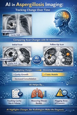
The role of AI in imaging follow-up for aspergillosis
Aspergillosis is not a single disease, but a spectrum of related conditions, including allergic bronchopulmonary aspergillosis (ABPA), chronic pulmonary aspergillosis (CPA), aspergilloma, subacute invasive aspergillosis, and Aspergillus bronchitis. Across this spectrum, imaging plays a central role in assessment, monitoring, and treatment planning.
Artificial intelligence (AI) is increasingly being explored in chest imaging as a supportive tool, particularly in the follow-up of chronic and complex lung disease. Its potential value in aspergillosis lies not in making diagnoses, but in consistent, data-driven image comparison.
Why aspergillosis is challenging for imaging interpretation
Imaging findings in aspergillosis are often:
-
Non-specific, overlapping with tuberculosis, non-tuberculous mycobacterial lung disease, lung cancer, vasculitis, and post-infectious scarring
-
Heterogeneous, even within the same patient
-
Slowly evolving, making change difficult to judge by eye
-
Mixed with other lung disease, such as bronchiectasis, asthma, fibrosis, or emphysema
For these reasons, imaging must always be interpreted in full clinical context and often benefits from specialist multidisciplinary discussion.
Where AI may add particular value
AI does not “know” that it is looking at aspergillosis. Instead, it compares imaging data directly, without assumptions about diagnosis. This can be helpful in several areas relevant to aspergillosis:
1. Consistent comparison of serial scans
AI can compare computed tomography (CT) scans over time using the same criteria each time, helping to:
-
Detect subtle interval change in cavities or nodules
-
Measure changes in cavity size, wall thickness, or internal content
-
Identify progression or stability that may be difficult to judge visually
This is particularly useful in chronic pulmonary aspergillosis, where progression may be slow and subtle.
2. Objective measurement of disease burden
AI can assist with:
-
Quantifying cavity volume or consolidation
-
Measuring extent of bronchiectasis or mucus plugging
-
Tracking changes following antifungal treatment or airway clearance
Objective measurements may help reduce subjectivity when monitoring response to treatment.
3. Highlighting areas for closer review
AI systems can flag areas of change or abnormality for radiologist attention. This may act as a “second set of eyes”, particularly in busy services, but does not replace expert review.
What AI cannot do in aspergillosis
It is important to be clear about the limitations:
-
AI cannot diagnose aspergillosis
-
AI cannot distinguish colonisation from active disease
-
AI cannot integrate symptoms, immune status, serology, or microbiology
-
AI cannot judge clinical significance or treatment need
For example, a change in cavity appearance may reflect active disease, treatment response, bacterial infection, bleeding, or simple movement of intracavitary material. Only expert clinical interpretation can determine significance.
Why radiologist expertise remains essential
In aspergillosis, small imaging changes can have very different meanings depending on context. Radiologists bring:
-
Experience in recognising mimics and artefacts
-
Understanding of treatment-related change
-
Ability to communicate uncertainty and recommend next steps
-
Integration of imaging with wider clinical information
AI may improve consistency and sensitivity, but responsibility for interpretation and reporting remains with the radiologist.
A balanced way to think about AI in aspergillosis imaging
In aspergillosis, artificial intelligence is best viewed as a tool that highlights and measures change, rather than one that explains or diagnoses it. Its strength lies in consistency; its limitation lies in lack of clinical understanding.
Used appropriately, AI may support safer and more consistent follow-up for people living with aspergillosis, while expert radiology and specialist clinical care remain central to diagnosis and management.

Using Radiopaedia and Online Imaging Resources Safely: What Expert Patients and Non-Specialist Clinicians Need to Know
Online radiology education platforms such as Radiopaedia (see aspergillosis images here) have transformed access to medical knowledge. They provide high-quality explanations of imaging findings, annotated examples, and differential diagnoses that are invaluable for learning, teaching, and patient empowerment.
For expert patients living with long-term conditions, and for non-specialist clinicians working outside radiology, these resources can greatly improve understanding of scan reports and discussions with healthcare teams. However, it is important to understand what these tools can – and cannot – do.
Radiopaedia is an educational resource, not a diagnostic service
Radiopaedia is designed to teach pattern recognition and radiological reasoning, not to provide individual diagnoses. The cases shown are curated examples, often with classic features, and are presented without the full clinical complexity that accompanies real patients.
Real-world imaging interpretation requires integration of:
-
Clinical history and symptoms
-
Laboratory results (for example inflammatory markers, microbiology, immunology)
-
Prior imaging and disease progression
-
Treatment history and response
-
Knowledge of common mimics and incidental findings
This clinical synthesis cannot be replicated by reviewing example images alone.
Why expert radiologist review still matters
For many diagnoses, there is no substitute for a radiologist formally reviewing and interpreting the imaging.
This is particularly true when:
-
Findings are subtle or evolving
-
Multiple conditions coexist (for example bronchiectasis, infection, scarring, and inflammation together)
-
Imaging appearances overlap between diseases
-
Treatment decisions depend on small but important changes over time
Radiologists are trained to recognise not only “textbook” appearances, but also atypical, incomplete, or misleading patterns, and to weigh uncertainty appropriately in their reports.
Imaging patterns are rarely diagnostic in isolation
Many imaging features are non-specific. For example:
-
Cavities can be caused by infection, inflammation, malignancy, or prior disease
-
Nodules may represent infection, scarring, inflammation, or benign change
-
Mucus plugging can occur in asthma, infection, allergic disease, or chronic airway disease
Educational resources often present differential diagnoses clearly, but deciding which diagnosis applies to a specific patient requires clinical judgment and experience.
A particular note for chronic lung and fungal disease
In complex conditions such as chronic lung disease, allergic lung disease, or fungal infections, imaging interpretation is especially nuanced. Appearances may change slowly, fluctuate with treatment, or overlap with other long-standing abnormalities.
Small changes that are significant to a specialist team may appear minor or ambiguous when viewed without context. Conversely, dramatic-looking findings may represent stable or inactive disease.
This is why specialist radiology input, often alongside multidisciplinary discussion, remains essential.
How expert patients and clinicians should use Radiopaedia
Used appropriately, Radiopaedia can:
-
Improve understanding of scan terminology
-
Help frame informed questions for clinicians
-
Support education and shared decision-making
-
Aid non-specialists in recognising when further advice is needed
It should not be used to:
-
Self-diagnose based on image similarity
-
Override formal radiology reports
-
Draw conclusions without clinical correlation
The key message
Radiopaedia and similar platforms are powerful educational tools. They enhance knowledge, confidence, and communication. But for many diagnoses, they complement rather than replace expert radiologist assessment.
The safest and most effective approach is to use educational resources alongside formal imaging reports, specialist input, and clinical discussion — not instead of them.
Understanding Aspergillosis Through Imaging: A Guide for Patients and Non-Specialist Clinicians
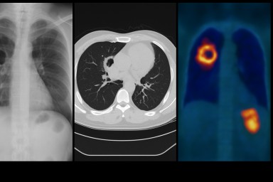
Imaging — especially chest X-ray and high-resolution CT (HRCT) — is one of the most important tools for recognising, diagnosing and monitoring aspergillosis. Because the condition can affect the lungs in very different ways, seeing what is happening inside the chest is essential for both patients and clinicians.
This guide explains why imaging matters, how it is used, and provides links to trusted resources that show what aspergillosis looks like on scans.
Why Imaging Matters in Aspergillosis
Aspergillosis affects the lungs deep within the airway and lung tissue. Many of these changes cannot be detected by a stethoscope or blood tests alone. Imaging helps detect:
-
mucus plugging
-
bronchiectasis (damaged widened airways)
-
cavities (holes) in the lungs
-
fungal balls (aspergillomas)
-
inflammation and consolidation
-
scarring or fibrosis
-
signs of haemoptysis risk
In Allergic Bronchopulmonary Aspergillosis (ABPA), imaging may show mucus impaction or central bronchiectasis.
In Chronic Pulmonary Aspergillosis (CPA), it may show cavities, thickened cavity walls, or a fungus ball.
In invasive aspergillosis, imaging can detect early nodules, the “halo sign”, or rapidly progressing changes.
Understanding these patterns helps clinicians choose the right treatment at the right time, and helps patients make sense of what is happening in their lungs.
Key Online Resources for Aspergillosis Imaging
Radiopaedia – Open Radiology Reference
One of the clearest, most comprehensive imaging resources available.
-
Pulmonary aspergillosis overview:
https://radiopaedia.org/articles/pulmonary-aspergillosis?lang=gb -
Invasive pulmonary aspergillosis:
https://radiopaedia.org/articles/invasive-pulmonary-aspergillosis?lang=gb -
Aspergilloma (fungal ball):
https://radiopaedia.org/articles/aspergilloma?lang=gb
Why it’s useful:
Radiopaedia shows real CT and X-ray examples from multiple patients, helping you understand what radiologists look for and how different forms of aspergillosis appear on imaging.
Peer-Reviewed Pictorial Reviews
These academic reviews provide side-by-side CT examples of the full spectrum of disease, written in an accessible style and extremely useful for both patients and clinicians.
-
Imaging Spectrum in Chronic Pulmonary Aspergillosis (Garg et al., 2022)
https://pmc.ncbi.nlm.nih.gov/articles/PMC9833062/ -
Pictorial Essay: Forms of Pulmonary Aspergillosis (Tamkevičiūtė et al., 2024)
(abstract) https://www.ejradiology.com/article/S0720-048X%2824%2900006-8/abstract
These explain in pictures:
-
how cavities form
-
how aspergillomas look
-
early vs late changes
-
how ABPA patterns differ from CPA
-
what stable vs progressive disease looks like
They are especially helpful for GPs, respiratory trainees, and nurses learning to interpret fungal disease imaging.
Classic RSNA Radiographics Review (Franquet et al.)
A foundational article describing radiologic patterns in allergic, chronic and invasive aspergillosis.
Although technical, this remains a reference standard for understanding how fungal disease presents on CT.
National Aspergillosis Centre (UK) – Patient-Friendly Information
Clear explanations of why imaging is needed, how CT is used, and what typical findings mean.
-
Chronic Pulmonary Aspergillosis (CPA):
https://mft.nhs.uk/wythenshawe/services/infectious-diseases/national-aspergillosis-centre/about-aspergillosis/chronic-pulmonary-aspergillosis-cpa/
These pages are ideal for newly diagnosed patients, people preparing for CT scans, and clinicians who want a quick overview.
Asthma + Lung UK – General Aspergillosis Overview
Patient-friendly explanations of the different types of aspergillosis, diagnosis and treatment.
Includes mention of imaging when relevant.
How Imaging Guides Clinical Decisions
1. Confirming the Diagnosis
Different forms of aspergillosis look different on scans:
-
ABPA may show “finger-in-glove” mucus plugging or central bronchiectasis.
-
CPA requires a cavity seen on imaging for >3 months.
-
Aspergilloma is a fungal ball sitting within a cavity.
-
Invasive disease shows nodules, halo signs, or rapidly evolving infiltrates.
Without CT imaging, these distinctions cannot reliably be made.
2. Checking for Complications
Imaging detects:
-
new cavities
-
cavity wall thickening
-
risk of haemoptysis (bleeding)
-
pleural thickening
-
fibrosis
-
co-existing infections
These findings often trigger urgent or proactive treatment modifications.
3. Monitoring Progression and Treatment Response
Symptoms alone are not enough. Imaging shows whether:
-
disease is stable or progressing
-
antifungal treatment is working
-
inflammation is reducing
-
mucus plugging is clearing
-
new areas are becoming involved
This is why people with CPA or ABPA may have repeat CT scans, usually every 6–12 months or when symptoms worsen.
Using These Resources Safely
These resources are not for self-diagnosis. Instead, they help:
-
patients understand their own scan reports
-
GPs recognise when to refer to a specialist
-
hospital teams spot aspergillosis in complex or unclear cases
-
nurses and allied health professionals visualise lung changes
-
clinicians communicate findings more clearly
Imaging must always be interpreted by a trained radiologist or specialist team, but these materials help demystify the process.
Summary
Imaging is central to the diagnosis and management of aspergillosis. Whether you are a patient trying to understand your scans, a GP seeing a complex chest X-ray, or a hospital clinician assessing breathlessness, these imaging resources provide clear, trustworthy examples.
By learning what different patterns look like — mucus plugging, cavities, aspergillomas, fibrosis or invasive changes — non-specialists can make more confident decisions, and patients can better understand their condition.