

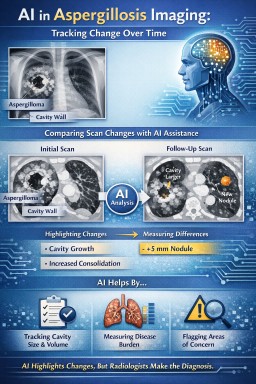
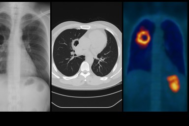
Medical imaging is an important part of diagnosing and monitoring many forms of aspergillosis. Chest X-rays, CT and high-resolution CT (HRCT) can show changes in the lungs such as cavities, fungal balls, bronchiectasis, fibrosis and other patterns that may help doctors understand what is happening.
Scans are only one part of the diagnosis. Imaging findings usually need to be interpreted alongside symptoms, previous scans, blood tests, microbiology and other clinical information. Different lung conditions can sometimes produce similar appearances on a scan.
This hub brings together more detailed information about CT, HRCT, X-ray and other imaging used in aspergillosis, including how scans are interpreted and how imaging can be used to follow changes over time.
Can Lung Cavities Heal in Aspergillosis? – Why some lung cavities shrink, why others remain, and how doctors assess treatment success in chronic pulmonary aspergillosis (CPA).
Topics in This Hub
Latest Imaging Articles



Related Hubs
Did this page help you?
Tell us in one minute